Bassett Collection of Stereoscopic Images of Human Anatomy
Pectoral region and axilla
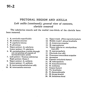
Left axilla (continued); general view of contents, clavicle removed
Image #91-2
KEYWORDS: Axilla, Overview.
Creative Commons
Stanford holds the copyright to the David L. Bassett anatomical images and has assigned Creative Commons license Attribution-Share Alike 4.0 International to all of the images.

For additional information regarding use and permissions, please contact the Medical History Center.
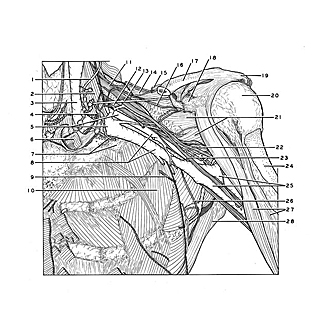
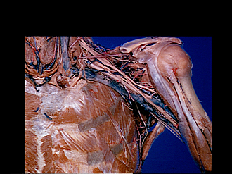
Pectoral region and axilla
Left axilla (continued); general view of contents, clavicle removed
The subclavius muscle and the medial two-thirds of the clavicle have been removed.
- Superficial cervical artery
- Anterior scalene muscle
- Internal jugular vein
- Phrenic nerve
- Upper pointer: Subclavian artery Lower pointer: Subclavian vein
- Sternohyoid muscle (visible through cut-out portion of capsule of sternoclavicular joint)
- Left pointer: Subclavian trunk Right pointer: Rib I
- Upper pointer: Axillary artery Lower pointer: Axillary vein
- Intercostobrachial nerve (cut off)
- External intercostal muscle
- Upper trunk
- Middle trunk (11 and 12 pertain to the supraclavicular part of the brachial plexus)
- Transverse scapular artery
- Supraspinatus muscle
- Inferior belly of omohyoid muscle (cut off)
- Suprascapular nerve
- Clavicle (cut off)
- Coracoid process of scapula
- Acromion
- Joint capsule of humerus
- Subscapularis muscle
- Musculocutaneous nerve
- Median nerve
- Body of humerus
- Brachial veins
- Thoracodorsal nerve
- Biceps brachii muscle
- Latissimus dorsi muscle