Bassett Collection of Stereoscopic Images of Human Anatomy
Pectoral region and axilla
Left sternoclavicular joint opened
Image #91-1
KEYWORDS: Fascia ligaments and tendons, Muscles and tendons, Pectoral region.
Creative Commons
Stanford holds the copyright to the David L. Bassett anatomical images and has assigned Creative Commons license Attribution-Share Alike 4.0 International to all of the images.

For additional information regarding use and permissions, please contact the Medical History Center.

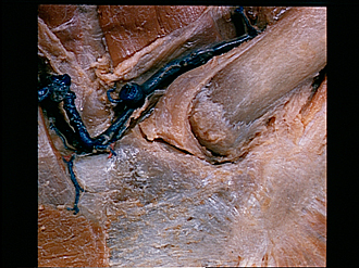
Pectoral region and axilla
Left sternoclavicular joint opened
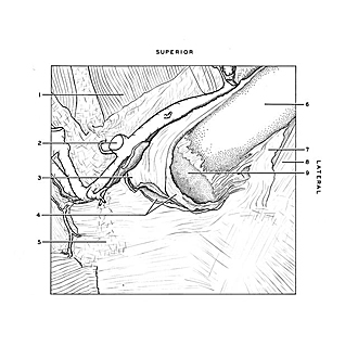
The capsule has been resected to expose the articular disc (4) which separates the medial and lateral cavities of the joint. The sternal end of the clavicle is covered with fibrocartilage. The unevenness of this surface is of common occurrence.
- Sternohyoid muscle
- Anterior jugular vein (cut off)
- Sternoclavicular joint capsule (pointer on interclavicular ligament which has been divided)
- Left pointer: Medial joint cavity Right pointer: Articular disc
- Manubrium of sternum
- Clavicle
- Costoclavicular ligament
- Tendon of origin of subclavius muscle
- Sternal articular surface clavicle (covered with fibrocartilage)