
Bassett Collection of Stereoscopic Images of Human Anatomy
Pectoral region and axilla
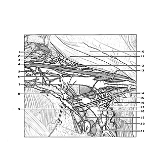
Left axilla (continued); relations of structures in posterior part of axillary fossa
Image #90-5
KEYWORDS: Axilla, Fascia ligaments and tendons, Neuralnetwork, Peripheral nervous system, Vasculature.
Creative Commons
Stanford holds the copyright to the David L. Bassett anatomical images and has assigned Creative Commons license Attribution-Share Alike 4.0 International to all of the images.

For additional information regarding use and permissions, please contact the Medical History Center.

Pectoral region and axilla
Left axilla (continued); relations of structures in posterior part of axillary fossa
The structures in the posterior part of the axilla have been exposed and are shown in relation to vessels and nerves already encountered. A distinct fascial lamina (21) is present in the cleft between the serratus anterior and subscapular muscles. Each of these muscles is also covered by its own fascia.
- Suprascapular nerve
- Subscapular nerve
- Posterior cord of brachial plexus
- Lateral cord of brachial plexus
- Anterior lateral thoracic nerve
- Upper pointer: Thoracoacromial artery (cut off) Lower pointer: Anterior medial thoracic nerve
- Upper pointer: Communicating loop between medial and lateral anterior thoracic nerves Lower pointer: Supreme thoracic artery
- Axillary artery and vein
- Intercostobrachial nerve
- Biceps brachii muscle (short head)
- Axillary nerve
- Musculocutaneous nerve
- Medial and lateral heads of median nerve
- Lateral thoracic artery
- Lymph vessel
- Upper pointer: Subscapular artery (accompanied by subscapular vein) Lower pointer: Teres major muscle (covered by fascia)
- Thoracodorsal nerve
- Axillary lymph nodes (lateral group)
- Long thoracic nerve
- Latissimus dorsi muscle
- Fascial layer (see text above)