Bassett Collection of Stereoscopic Images of Human Anatomy
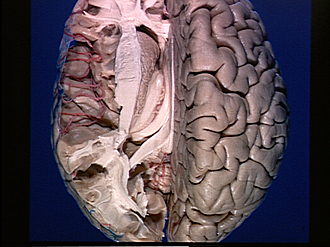
Exploration of the brain from its superior aspect
Caudate nucleus; lamina affixa; medullary substance of insula
Image #9-6
KEYWORDS: Brain, Parietal lobe, Telencephalon, Vasculature.
Creative Commons
Stanford holds the copyright to the David L. Bassett anatomical images and has assigned Creative Commons license Attribution-Share Alike 4.0 International to all of the images.

For additional information regarding use and permissions, please contact the Medical History Center.


Exploration of the brain from its superior aspect
Caudate nucleus; lamina affixa; medullary substance of insula
The cortex of the upper half of the insula has been scraped away to reveal the underlying medullary substance. The ependymal layer which covered the caudate nucleus has been removed. Much of the choroid plexus in the central part of the lateral ventricle has been cut away to expose its attachment to the lips (taeniae) of the choroidal fissure (cleft between fornix and lamina affixa). Note the choroidal artery (a branch of the a. cerebri posterior) passing anteriorly in this region.
- Medullary substance projecting into superior frontal gyrus
- Head of caudate nucleus (ependyma removed)
- Medullary substance of insula
- Internal capsule
- Anterior tubercle of thalamus
- Great anastomotic vein (cut end)
- Lamina affixa
- Choroidal fissure (cleft between taenia chorioidea and head of fornix)
- Fornix (crus)
- Cut end of superior longitudinal fasciculus
- Hippocampus
- Calcar avis
- Occipital part radiations of corpus callosum
- Lingual gyrus (cut across)
- Cingulum (divided)
- Precentral gyrus
- Central sulcus
- Branch of posterior cerebral artery in parieto-occipital fissure
- Interparietal sulcus