Bassett Collection of Stereoscopic Images of Human Anatomy
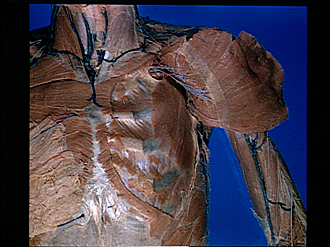
Pectoral region and axilla
Nerve and blood supply of left pectoralis major muscle
Image #89-3
KEYWORDS: Forearm, Neuralnetwork, Pectoral region, Peripheral nervous system, Vasculature.
Creative Commons
Stanford holds the copyright to the David L. Bassett anatomical images and has assigned Creative Commons license Attribution-Share Alike 4.0 International to all of the images.

For additional information regarding use and permissions, please contact the Medical History Center.


Pectoral region and axilla
Nerve and blood supply of left pectoralis major muscle
The pectoralis major (9) has been reflected laterally from its clavicular (2) and sternocostal (3) origins. A narrow band of the pectoral fascia (8) which invested the deep surface of the muscle has been retained.
- Sternocleidomastoid muscle
- Clavicular part pectoralis major muscle (cut across)
- Sternocostal part pectoralis major muscle (cut across)
- Upper pointer: Abdominal part pectoralis major muscle (cut across) Lower pointer: Sheath of rectus abdominis muscle
- Supraclavicular nerves
- Axillary vein
- Anterior thoracic nerve (branches of thoracoacromial vessels lie close to nerve)
- Remnant of pectoral fascia
- Pectoralis major muscle (reflected laterally)
- Pectoralis minor muscle
- Rib II
- Body of sternum
- Pectoral fascia (at former position of lateral border of pectoralis major)
- Serratus anterior muscle
- Lateral cutaneous branch of intercostal nerve VI