Bassett Collection of Stereoscopic Images of Human Anatomy
Osteology
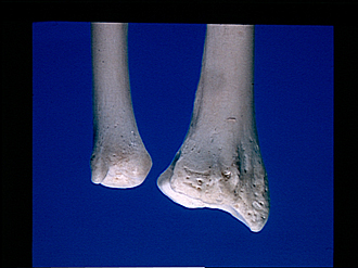
Right radius and ulna, posterior view of distal parts
Image #87-1
KEYWORDS: Forearm, Muscles and tendons.
Creative Commons
Stanford holds the copyright to the David L. Bassett anatomical images and has assigned Creative Commons license Attribution-Share Alike 4.0 International to all of the images.

For additional information regarding use and permissions, please contact the Medical History Center.


Osteology
Right radius and ulna, posterior view of distal parts
- Ulna
- Dorsal surface
- Head (pointer on groove for tendon of extensor carpi ulnaris muscle)
- Styloid process
- Articular circumference
- Radius
- Ulnar notch (of radius)
- Carpal articular surface
- Dorsal surface
- Groove for tendons of common extensor digitorum muscle and extensor indicis muscle
- Upper pointer: Groove for tendon of extensor pollicis longus muscle Lower pointer: Dorsal tubercle
- Upper pointer: Groove for tendon of extensor carpi radialis brevis muscle Lower pointer: Groove for tendon of extensor carpi radialis longus muscle
- Styloid process