Bassett Collection of Stereoscopic Images of Human Anatomy
Dissection of head and neck from a posterior approach
Pharynx and esophagus; posterior aspect of larynx
Image #82-6
KEYWORDS: Esophagus, Pharynx, Throat.
Creative Commons
Stanford holds the copyright to the David L. Bassett anatomical images and has assigned Creative Commons license Attribution-Share Alike 4.0 International to all of the images.

For additional information regarding use and permissions, please contact the Medical History Center.
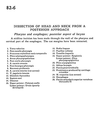
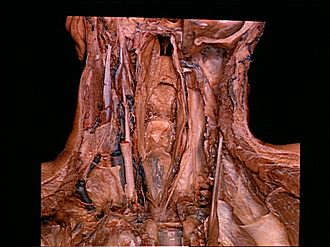
Dissection of head and neck from a posterior approach
Pharynx and esophagus; posterior aspect of larynx
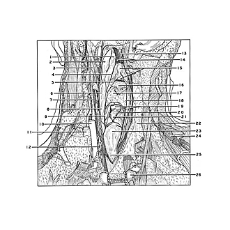
A midline incision has been made through the wall of the pharynx and cervical part of the esophagus. The cut margins have been retracted.
- Torus tubarius
- Nasal part pharynx
- Styloid process temporal bone
- Salpingopharyngeal fold
- Palatopharyngeal arch
- Oral part pharynx
- External carotid artery
- Laryngeal part of pharynx
- Piriform recess
- Internal carotid artery (cut across)
- Internal jugular vein
- Thyroid gland
- Nasal septum
- Choanae
- Upper pointer: Soft palate Lower pointer: Uvula (poorly developed)
- Root of tongue
- Vallate papillae
- Lingual tonsil
- Upper pointer: Epiglottis Lower pointer: Pharyngoepiglottic fold
- Aryepiglottic fold
- Laryngeal ventricle
- Prevertebral fascia
- Prominence produced by cricoid cartilage
- Trapezius muscle (cut across)
- Esophagus
- Superior articular surface thoracic vertebrae II