Bassett Collection of Stereoscopic Images of Human Anatomy
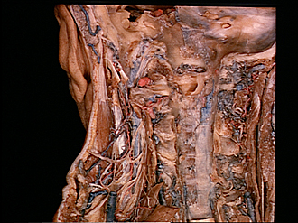
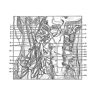
Dissection of head and neck from a posterior approach
Relations of left sternocleidomastoid muscle, internal jugular vein, cervical plexus and cervical spine
Image #80-7
KEYWORDS: Bones cartilage joints, Cervical vertebrae, Muscles and tendons, Peripheral nervous system, Vasculature.
Creative Commons
Stanford holds the copyright to the David L. Bassett anatomical images and has assigned Creative Commons license Attribution-Share Alike 4.0 International to all of the images.

For additional information regarding use and permissions, please contact the Medical History Center.

Dissection of head and neck from a posterior approach
Relations of left sternocleidomastoid muscle, internal jugular vein, cervical plexus and cervical spine
The fascia has been removed from the upper part of the sternocleidomastoid muscle, which has been pulled somewhat anteriorly. The bodies of the upper cervical vertebrae have been exposed to the left of the midline. The levator scapulae muscle has been cut from its origins and retracted medially and inferiorly to expose the cervical plexus.
- Internal jugular vein (pointer at inferior margin of jugular foramen)
- Hypoglossal nerve (XII)
- Accessory nerve (XI)
- Tendon of insertion of longissimus capitis muscle (divided by occipital artery)
- Splenius capitis muscle (cut across)
- Anterior branch of cervical nerve I
- Posterior belly of digastric muscle
- Levator scapulae muscle (cut across; a similar, more inferior origin is visible medial to the third cervical nerve (24))
- Sternocleidomastoid muscle
- Ascending cervical artery
- Lesser occipital nerve
- Upper pointer: Greater auricular nerve and superficial cutaneous nerve Lower pointer: Supraclavicular nerves
- Posterior cervical triangle
- External jugular vein (cut across)
- Upper pointer: Foramen magnum (anterior margin) Lower pointer: Dens (axis)
- Right alar ligament
- Joint cavity between dens and transverse ligament of atlas
- Inferior oblique capitis muscle (cut across)
- Cervical nerve III
- Upper pointer: Arch of axis Lower pointer: Roots of cervical nerve III
- Semispinalis capitis muscle
- Splenius capitis muscle
- Upper pointer: Basivertebral vein (cut off at junction with longitudinal vertebral sinus) Lower pointer: Body cervical vertebra llI
- Cervical nerve III
- Intervertebral joint (between articular processes of C. III- IV)
- Ganglion cervical nerve V
- Annulus fibrosus intervertebral disc (C. IV-V)
- Articular surface superior cervical vertebra VI
- Levator scapulae muscle (also see 8 above)