Bassett Collection of Stereoscopic Images of Human Anatomy
Dissection of head and neck from a posterior approach
Cruciate ligament; alar ligaments
Image #80-4
KEYWORDS: Bones cartilage joints, Connective tissue, Cervical vertebrae, Fascia and connective tissue.
Creative Commons
Stanford holds the copyright to the David L. Bassett anatomical images and has assigned Creative Commons license Attribution-Share Alike 4.0 International to all of the images.

For additional information regarding use and permissions, please contact the Medical History Center.
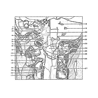
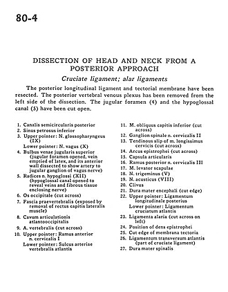
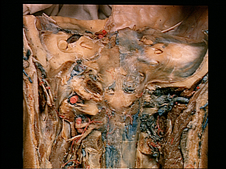
Dissection of head and neck from a posterior approach
Cruciate ligament; alar ligaments
The posterior longitudinal ligament and tectorial membrane have been resected. The posterior vertebral venous plexus has been removed from the left side of the dissection. The jugular foramen (4) and the hypoglossal canal (5) have been cut open.
- Posterior semicircular canal
- Inferior petrosal sinus
- Upper pointer: Glossopharyngeal nerve (IX) Lower pointer: Vagus nerve (X)
- Superior jugular venous bulb (jugular foramen opened, vein emptied of latex, and its anterior wall dissected to show artery to jugular ganglion of vagus nerve)
- Roots of hypoglossal nerve (XII) (hypoglossal canal opened to reveal veins and fibrous tissue enclosing nerve)
- Occipital bone (cut across)
- Prevertebral fascia (exposed by removal of rectus capitis lateralis muscle)
- Atlantooccipital cavity of articulation
- Vertebral artery (cut across)
- Upper pointer: Anterior branch of cervical nerve I Lower pointer: Groove in atlas for vertebral artery
- Inferior oblique capitis muscle (cut across)
- Spinal ganglion cervical nerve II
- Tendinous slip of longissimus cervicis muscle (cut across)
- Arch of axis (cut across)
- Joint capsule
- Posterior branch cervical nerve III
- Levator scapulae muscle
- Trigeminal nerve (V)
- Vestibulocochlear nerve (VIII)
- Clivus
- Dura mater encephali (cut edge)
- Upper pointer: Posterior longitudinal ligament Lower pointer: Cruciate ligament atias
- Alar ligament (cut across on left)
- Position of dens (axis)
- Cut edge of tectorial membrane
- Transverse ligament atlas (part of cruciate ligament)
- Dura mater