Bassett Collection of Stereoscopic Images of Human Anatomy
Exploration of the brain from its superior aspect
Superior longitudinal fasciculus
Image #8-7
KEYWORDS: Brain, Frontal lobe, Occipital lobe, Parietal lobe, Telencephalon, Temporal lobe, Overview.
Creative Commons
Stanford holds the copyright to the David L. Bassett anatomical images and has assigned Creative Commons license Attribution-Share Alike 4.0 International to all of the images.
For additional information regarding use and permissions, please contact the Medical History Center.
Exploration of the brain from its superior aspect
Superior longitudinal fasciculus
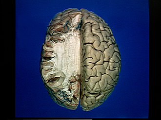
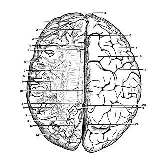
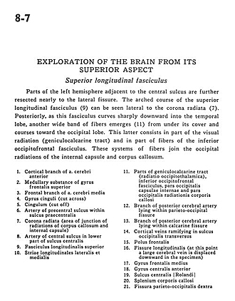
Parts of the left hemisphere adjacent to the central sulcus are further resected nearly to the lateral fissure. The arched course of the superior longitudinal fasciculus (9) can be seen lateral to the corona radiata (7). Posteriorly, as this fasciculus curves sharply downward into the temporal lobe, another wide band of fibers emerges (11) from under its cover and courses toward the occipital lobe. This latter consists in part of the visual radiation (geniculocalcarine tract) and in part of fibers of the inferior occipitofrontal fasciculus. These systems of fibers join the occipital radiations of the internal capsule and corpus callosum.
- Cortical branch of anterior cerebral artery
- Medullary substance of superior frontal gyrus
- Frontal branch of middle cerebral artery
- Cingulate gyrus (cut across)
- Cingulum (cut off)
- Artery of precentral sulcus within precentral sulcus
- Corona radiata (area of junction of radiations of corpus callosum and internal capsule)
- Artery of central sulcus in lower part of central sulcus
- Superior longitudinal fasciculus
- Lateral and medial longitudinal striae
- Parts of geniculocalcarine tract (optic radiations), inferior occipitofrontal fasciculus, occipital part internal capsule and occipital part of radiations of corpus callosum
- Branch of posterior cerebral artery lying within parieto-occipital fissure
- Branch of posterior cerebral artery lying within calcarine fissure
- Cortical veins ramifying in transverse occipital sulcus
- Frontal pole
- Longitudinal fissure (at this point a large cerebral vein is displaced downward in the specimen)
- Medial frontal gyrus
- Precentral gyrus
- Central sulcus (Rolandic)
- Corpus callosum (splenium)
- Parieto-occipital fissure right


