Bassett Collection of Stereoscopic Images of Human Anatomy
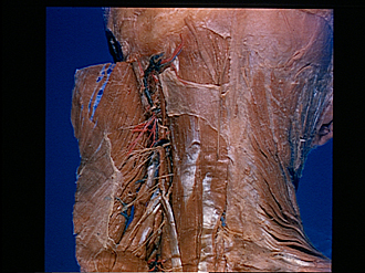
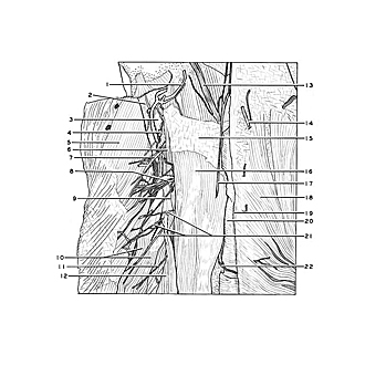
Dissection of head and neck from a posterior approach
Nerve supply to left splenius capitis and cervicis muscles
Image #79-2
KEYWORDS: Fascia and connective tissue, Muscles and tendons, Peripheral nervous system.
Creative Commons
Stanford holds the copyright to the David L. Bassett anatomical images and has assigned Creative Commons license Attribution-Share Alike 4.0 International to all of the images.

For additional information regarding use and permissions, please contact the Medical History Center.
Dissection of head and neck from a posterior approach
Nerve supply to left splenius capitis and cervicis muscles
The muscles have been cut from their spinal origins (the splenius capitis muscle also cut near its cranial insertion) and reflected laterally. A portion of the fascia which covered the deep surface of the splenius capitis muscle has been preserved. The fascia (15) of the semispinalis capitis muscle (16) has been retained only in a small area.
- Occipital artery
- Mastoid process
- Superior oblique capitis muscle
- Suboccipital lymph node
- Splenius capitis muscle (reflected)
- Aberrant muscular slip from longissimus capitis muscle to semispinalis capitis muscle
- Descending branch occipital artery
- Muscular branch of posterior branch cervical nerve II
- Longus capitis muscle
- Splenius cervicis muscle (reflected)
- Muscular branch of posterior branch cervical nerve IV
- Longissimus cervicis muscle
- Greater occipital nerve
- Third occipital nerve right
- Fascia of semispinalis capitis muscle
- Semispinalis capitis muscle
- Third occipital nerve left
- Trapezius muscle right
- Nuchal ligament
- Aponeurosis of origin of splenius capitis muscle (cut across)
- Muscular branches of posterior branch cervical nerve III
- Cutaneous branch of posterior branch cervical nerve IV