
Bassett Collection of Stereoscopic Images of Human Anatomy
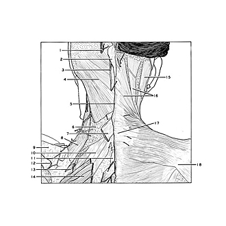
Dissection of head and neck from a posterior approach
Splenius capitis and levator scapulae muscles
Image #78-7
KEYWORDS: Connective tissue, Muscles and tendons, Fascia and connective tissue.
Creative Commons
Stanford holds the copyright to the David L. Bassett anatomical images and has assigned Creative Commons license Attribution-Share Alike 4.0 International to all of the images.

For additional information regarding use and permissions, please contact the Medical History Center.
Dissection of head and neck from a posterior approach
Splenius capitis and levator scapulae muscles
The left trapezius and sternocleidomastoid muscles have been removed. The rhomboideus major and minor muscles have been divided. The fascia of the splenius capitis muscle has been cut away with the exception of a band (6) which has been retained across the inferior part of the muscle.
- Greater occipital nerve
- Tendon of origin of trapezius muscle left
- Third occipital nerve
- Splenius capitis muscle
- Nuchal ligament
- Fascia of splenius capitis muscle
- Rhomboid minor muscle (cut across near origin)
- Levator scapulae muscle
- Supraspinatus fascia
- Posterior superior serratus muscle
- Rhomboid major muscle (aponeurosis of origin)
- Tendon of insertion of trapezius muscle into spine of scapula
- Rhomboid minor muscle (cut across near insertion)
- Rhomboid major muscle
- Sternocleidomastoid muscle
- Trapezius muscle
- Spinous process cervical vertebra VII
- Spine of scapula