
Bassett Collection of Stereoscopic Images of Human Anatomy
Dissection of anterior and lateral regions of neck
Larynx; vocal cord and vocal muscle; lateral and posterior cricoarytenoid muscles; left lateral view
Image #77-7
KEYWORDS: Muscles and tendons, Throat.
Creative Commons
Stanford holds the copyright to the David L. Bassett anatomical images and has assigned Creative Commons license Attribution-Share Alike 4.0 International to all of the images.

For additional information regarding use and permissions, please contact the Medical History Center.
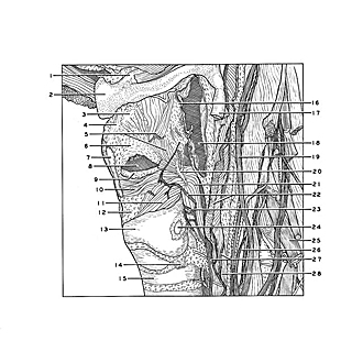

Dissection of anterior and lateral regions of neck
Larynx; vocal cord and vocal muscle; lateral and posterior cricoarytenoid muscles; left lateral view
The external part of the thyroarytenoid muscle has been removed and the internal part (vocal muscle) retained.
- Lesser horn hyoid bone
- Body hyoid bone
- Epiglottal cartilage
- Thyrohyoid ligament
- Upper pointer: Thyroepiglotticus muscle (cut across) Lower pointer: Aryepiglotticus muscle
- Quadrangular membrane
- Thyroid cartilage (cut across)
- Laryngeal ventricle (opened)
- Vocal cord
- Vocalis muscle
- Lateral cricoarytenoid muscle
- Conus elasticus
- Arch of cricoid cartilage
- Lateral suspensory ligament of thyroid gland (cut off)
- Trachea
- Aryepiglottic fold
- Superior horn thyroid cartilage (cut across)
- Piriform recess (opened)
- Vagus nerve (X)
- Arytenoid cartilage
- Posterior cricoarytenoid muscle
- Upper pointer: Anterior branch inferior laryngeal nerve Lower pointer: Inferior pharyngeal constrictor muscle (cut across)
- Posterior branch inferior laryngeal nerve
- Articular surface of thyroid
- Cervical nerve VI
- Vertebral artery
- Branches of recurrent esophageal nerve
- Esophagus