Bassett Collection of Stereoscopic Images of Human Anatomy
Dissection of anterior and lateral regions of neck
Relations of inferior cervical ganglion, anterior view
Image #76-4
KEYWORDS: Peripheral nervous system.
Creative Commons
Stanford holds the copyright to the David L. Bassett anatomical images and has assigned Creative Commons license Attribution-Share Alike 4.0 International to all of the images.

For additional information regarding use and permissions, please contact the Medical History Center.
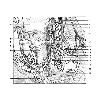
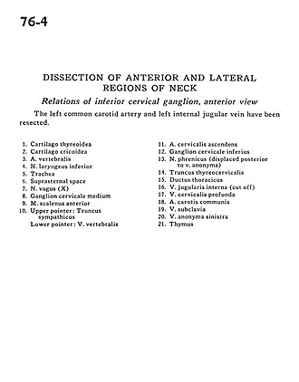
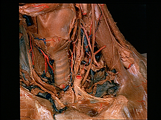
Dissection of anterior and lateral regions of neck
Relations of inferior cervical ganglion, anterior view
The left common carotid artery and left internal jugular vein have been resected.
- Thyroid cartilage
- Cricoid cartilage
- Vertebral artery
- Inferior laryngeal nerve
- Trachea
- Suprasternal space
- Vagus nerve (X)
- Middle cervical ganglion
- Anterior scalene muscle
- Upper pointer: Sympathetic trunk Lower pointer: Vertebral vein
- Ascending cervical artery
- Inferior cervical ganglion
- Phrenic nerve (displaced posterior to brachiocephalic vein)
- Thyrocervical trunk
- Thoracic duct
- Internal jugular vein (cut off)
- Deep cervical vein
- Common carotid artery
- Subclavian vein
- Brachiocephalic vein left
- Thymus