
Bassett Collection of Stereoscopic Images of Human Anatomy
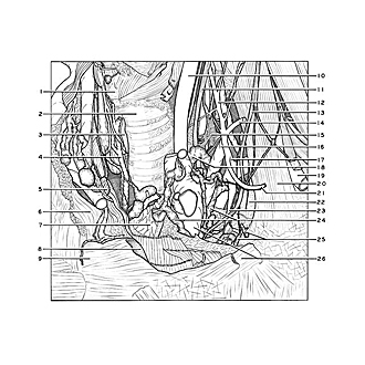
Dissection of anterior and lateral regions of neck
Thoracic duct; structures related to cupula of pleura, left anterolateral view
Image #76-3
KEYWORDS: Lymphatics, Vasculature.
Creative Commons
Stanford holds the copyright to the David L. Bassett anatomical images and has assigned Creative Commons license Attribution-Share Alike 4.0 International to all of the images.

For additional information regarding use and permissions, please contact the Medical History Center.
Dissection of anterior and lateral regions of neck
Thoracic duct; structures related to cupula of pleura, left anterolateral view
The internal jugular vein has been cut off and the left innominate vein retracted anteromedially.
- Cricoid cartilage
- Trachea
- Isthmus of thyroid gland (cut across)
- Inferior thyroid vein right
- Sternothyroid muscle
- Sternal extremity of clavicle (covered by joint capsule)
- Thymus
- Articular surface of manubrium of sternum for left clavicle
- Manubrium of sternum
- Common carotid artery
- Vagus nerve (X)
- Ascending cervical artery
- Phrenic nerve
- Superficial cervical artery
- Inferior cervical ganglion
- Vertebral vein
- Thyrocervical trunk
- Thoracic duct
- Subclavian artery
- Anterior scalene muscle
- Transverse scapular artery
- Cupula pleurae
- Internal thoracic (mammary) artery (note internal mammary nerve plexus)
- Anterior and posterior bronchomediastinal lymphatic trunks
- Left internal mammary lymphatic vessels
- Upper pointer: Anonymous vein left (retracted anteromedially) Lower pointer: Costoclavicular ligament