Bassett Collection of Stereoscopic Images of Human Anatomy
Dissection of anterior and lateral regions of neck
Levator scapulae and scalene muscles, lateral view
Image #75-6
KEYWORDS: Muscles and tendons.
Creative Commons
Stanford holds the copyright to the David L. Bassett anatomical images and has assigned Creative Commons license Attribution-Share Alike 4.0 International to all of the images.

For additional information regarding use and permissions, please contact the Medical History Center.
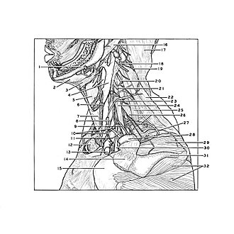
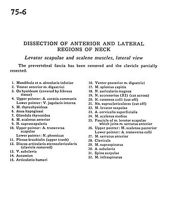
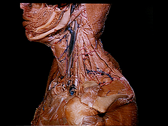
Dissection of anterior and lateral regions of neck
Levator scapulae and scalene muscles, lateral view
The prevertebral fascia has been removed and the clavicle partially resected.
- Mandible and inferior alveolar nerve
- Anterior belly digastric muscle
- Hyoid bone (covered by fibrous tissue)
- Upper pointer: Common carotid artery Lower pointer: Internal jugular vein
- Thyrohyoid muscle
- Ansa hypoglossi
- Thyroid gland
- Anterior scalene muscle
- Suprascapular nerve
- Upper pointer: Transverse scapular artery Lower pointer: Phrenic nerve
- Brachial plexus (upper trunk)
- Sternoclavicular articular disc (clavicle removed)
- Subclavian vein
- Acromion
- Glenohumeral joint
- Posterior belly of digastric muscle
- Splenius capitis muscle
- Greater auricular nerve
- Accessory nerve (XI) (cut across)
- Superficial cutaneous nerve (cut off)
- Supraclavicular nerves (cut off)
- Levator scapulae muscle
- Superficial cervical artery
- Middle scalene muscle
- Fascicle of levator scapulae muscle which joins serratus anterior muscle
- Upper pointer: Posterior scalene muscle Lower pointer: transverse colli artery
- Serratus anterior muscle
- Clavicle
- Supraspinatus muscle
- Subclavian artery
- Infraspinatus muscle