
Bassett Collection of Stereoscopic Images of Human Anatomy
Dissection of anterior and lateral regions of neck
Deep layers of infrahyoid muscles, anterior view
Image #74-4
KEYWORDS: Muscles and tendons, Throat.
Creative Commons
Stanford holds the copyright to the David L. Bassett anatomical images and has assigned Creative Commons license Attribution-Share Alike 4.0 International to all of the images.

For additional information regarding use and permissions, please contact the Medical History Center.

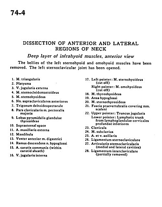
Dissection of anterior and lateral regions of neck
Deep layers of infrahyoid muscles, anterior view
The bellies of the left sternohyoid and omohyoid muscles have been removed. The left sternoclavicular joint has been opened.
- Depressor anguli oris muscle
- Platysma
- External jugular vein
- Sternocleidomastoid muscle
- Sternohyoid muscle
- Anterior supraclavicular nerves
- Deltopectoral triangle
- Clavicular part pectoralis major muscle
- Pyramidal lobe of thyroid gland
- Suprasternal space
- External maxillary artery
- Mandible
- Anterior belly digastric muscle
- Descending branch hypoglossal nerve
- Common carotid artery (within carotid sheath)
- Internal jugular vein
- Left pointer: Sternohyoid muscle (cut off) Right pointer: Omohyoid muscle (cut off)
- Thyrohyoid muscle
- Ansa hypoglossi
- Sternothyroid muscle
- Prevertebral fascia covering scalene muscles
- Upper pointer: Jugular trunk Lower pointer: Lymphatic trunk from inferior deep cervical lymph nodes
- Clavicle
- Subclavius muscle
- Axillary artery and vein
- Sternoclavicular ligament
- Sternoclavicular joint (medial and lateral cavities)
- Interclavicular ligament (partially removed)