Bassett Collection of Stereoscopic Images of Human Anatomy
Dissection of anterior and lateral regions of neck
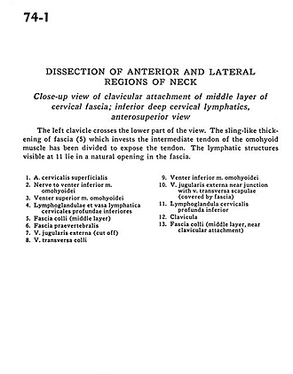
Close-up view of clavicular attachment of middle layer of cervical fascia; inferior deep cervical lymphatics, anterosuperior view
Image #74-1
KEYWORDS: Fascia and connective tissue, Lymphatics, Muscles and tendons.
Creative Commons
Stanford holds the copyright to the David L. Bassett anatomical images and has assigned Creative Commons license Attribution-Share Alike 4.0 International to all of the images.

For additional information regarding use and permissions, please contact the Medical History Center.
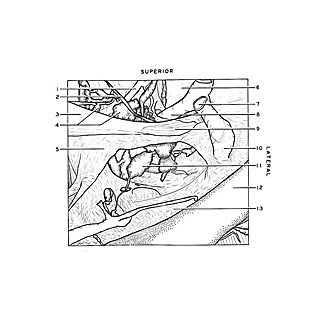
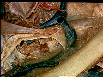
Dissection of anterior and lateral regions of neck
Close-up view of clavicular attachment of middle layer of cervical fascia; inferior deep cervical lymphatics, anterosuperior view
The left clavicle crosses the lower part of the view. The sling-like thickening of fascia (5) which invests the intermediate tendon of the omohyoid muscle has been divided to expose the tendon. The lympathic structures visible at 11 lie in a natural opening in the fascia.
- Superficial cervical artery
- Nerve to inferior belly omohyoid muscle
- Superior belly omohyoid muscle
- Inferior deep cervical lymph nodes and lymph vessels
- Superficial fascia (middle layer)
- Prevertebral fascia
- External jugular vein (cut off)
- Superficial transverse vein
- Inferior belly omohyoid muscle
- External jugular vein near junction with transverse scapular vein (covered by fascia)
- Inferior deep cervical lymph node
- Clavicle
- Superficial fascia (middle layer, near clavicular attachment)