Bassett Collection of Stereoscopic Images of Human Anatomy
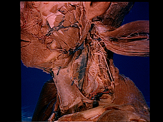
Dissection of anterior and lateral regions of neck
Deep cervical lymphatic nodes and vessels, left lateral view
Image #73-5
KEYWORDS: Lymphatics.
Creative Commons
Stanford holds the copyright to the David L. Bassett anatomical images and has assigned Creative Commons license Attribution-Share Alike 4.0 International to all of the images.

For additional information regarding use and permissions, please contact the Medical History Center.


Dissection of anterior and lateral regions of neck
Deep cervical lymphatic nodes and vessels, left lateral view
The sternocleidomastoid muscle has been retracted posteriorly. The external layer of cervical fascia and the parotideomasseteric fascia have been removed.
- Parotid gland
- Infraorbital nerve
- Buccinator muscle
- Masseter muscle
- External maxillary artery
- Superficial branch facial nerve (cut off)
- Mandible (periosteum intact)
- Submandibular gland
- Superior deep cervical lymph nodes
- Ansa hypoglossi
- Omohyoid muscle
- Superficial cervical artery
- Posterior auricular lymph nodes
- Greater auricular nerve
- Superior deep cervical lymph nodes
- Accessory nerve (XI) entering sternocleidomastoid muscle (note communication from cervical nerve III)
- Sternocleidomastoid muscle
- Cervical nerve IIl
- Superficial cutaneous nerve (cut off)
- Accessory nerve (XI)
- Internal jugular vein (within carotid sheath)
- Cervical nerve IV
- Superficial cervical lymph node
- Trapezius muscle
- Prevertebral fascia