
Bassett Collection of Stereoscopic Images of Human Anatomy
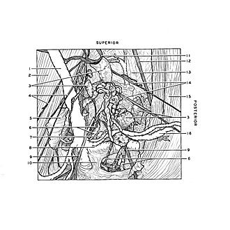
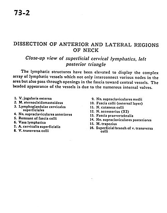
Dissection of anterior and lateral regions of neck
Close-up view of superficial cervical lymphatics, left posterior triangle
Image #73-2
KEYWORDS: Lymphatics.
Creative Commons
Stanford holds the copyright to the David L. Bassett anatomical images and has assigned Creative Commons license Attribution-Share Alike 4.0 International to all of the images.

For additional information regarding use and permissions, please contact the Medical History Center.
Dissection of anterior and lateral regions of neck
Close-up view of superficial cervical lymphatics, left posterior triangle
The lymphatic structures have been elevated to display the complex array of lymphatic vessels which not only interconnect various nodes in the area but also pass through openings in the fascia toward central vessels. The beaded appearance of the vessels is due to the numerous internal valves.
- External jugular vein
- Sternocleidomastoid muscle
- Superficial cervical lymph glands
- Anterior supraclavicular nerves
- Remnant of cervical fascia
- Lymphatic vessel
- Superficial cervical artery
- Transverse cervical vein
- Medial supraclavicular nerves
- Cervical fascia (external layer)
- Cutaneus colli nerve
- Accessory nerve (XI)
- Prevertebral fascia
- Posterior supraclavicular nerves
- Trapezius muscle
- Superficial branch of transverse cervical vein
- [Legend above restored translation from Latin]