
Bassett Collection of Stereoscopic Images of Human Anatomy
Dissection of nasal fossae, nasal pharynx, and palate
Blood vessels and nerves of nasal septum, left lateral view
Image #71-7
KEYWORDS: Bones cartilage joints, Face, Mouth, Nose, Peripheral nervous system, Vasculature.
Creative Commons
Stanford holds the copyright to the David L. Bassett anatomical images and has assigned Creative Commons license Attribution-Share Alike 4.0 International to all of the images.

For additional information regarding use and permissions, please contact the Medical History Center.
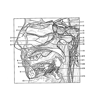
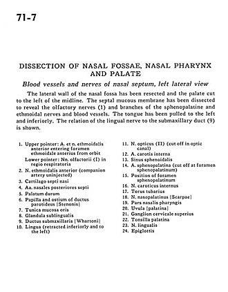
Dissection of nasal fossae, nasal pharynx, and palate
Blood vessels and nerves of nasal septum, left lateral view
The lateral wall of the nasal fossa has been resected and the palate cut to the left of the midline. The septal mucous membrane has been dissected to reveal the olfactory nerves (1) and branches of the sphenopalatine and ethmoidal nerves and blood vessels. The tongue has been pulled to the left and inferiorly. The relation of the lingual nerve to the submaxillary duct (9) is shown.
- Upper pointer: Anterior ethmoidal nerve and artery entering anterior ethmoidal foramen from orbit Lower pointer: Olfactory nerve (I) in respiratory region
- Anterior ethmoidal nerve (companion artery uninjected)
- Septal cartilage
- Posterior septal arteries
- Hard palate
- Papilla and ostium of parotid duct
- Oral mucosa
- Sublingual gland
- Submandibular duct
- Tongue (retracted inferiorly and to the left)
- Optic nerve (II) (cut off in optic canal)
- Internal carotid artery
- Sphenoid sinus
- Sphenopalatine artery (cut off at sphenopalatine foramen)
- Position of sphenopalatine foramen
- Internal carotid nerve
- Torus tubarius
- Nasopalatine nerve
- Nasal part pharynx
- Uvula (palatine)
- Superior cervical ganglion
- Palatine tonsil
- Lingual nerve
- Epiglottis