Bassett Collection of Stereoscopic Images of Human Anatomy
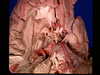
Exploration of the brain from its basal aspect
Transverse fissure and lateral ventricle exposed by removal of thalamus
Image #7-1
KEYWORDS: Brain, Diencephalon, Telencephalon, Ventricules.
Creative Commons
Stanford holds the copyright to the David L. Bassett anatomical images and has assigned Creative Commons license Attribution-Share Alike 4.0 International to all of the images.

For additional information regarding use and permissions, please contact the Medical History Center.
Exploration of the brain from its basal aspect
Transverse fissure and lateral ventricle exposed by removal of thalamus
The thalamus has now been cut away so that only its most medial and anterior portions remain. The lamina affixa (6), which covered the upper lateral part of the thalamus, is partially removed so that in effect the choroidal fissure (8) is opened from below and one looks directly into the medial part of the body of the lateral ventricle which is collapsed. The roof of the ventricle, formed by the corpus callosum, is seen at (7).
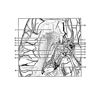
- Frontal part corona radiata
- Ependymal membrane of superior surface of caudate nucleus
- Superior occipitofrontal fasciculus
- Internal capsule (cut across at junction with corona radiata)
- Superior longitudinal fasciculus
- Lamina affixa (partially cut away)
- Corpus callosum (inferior surface visible through body of lateral ventricle)
- Choroid plexus lateral ventricle (attached to head of fornix)
- Choroid plexus inferior horn of lateral ventricle
- Longitudinal fissure (cerebral)
- Anterior communicating artery
- Anterior commissure
- Stria terminalis (note continuity with parolfactory area)
- Anterior perforated substance
- Mamillothalamic tract (Vicq d'Azyr)
- Mamillary body
- Fornix (body)
- Cerebral peduncle
- Dentate fascia (hippocampus)