
Bassett Collection of Stereoscopic Images of Human Anatomy
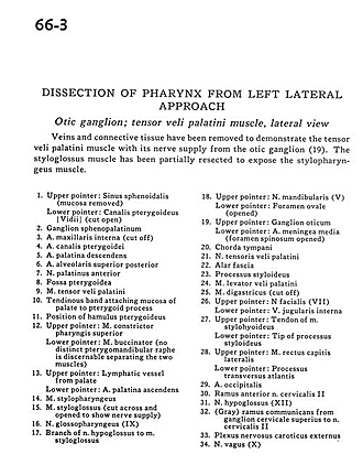
Dissection of pharynx from left lateral approach
Otic ganglion; tensor veli palatini muscle, lateral view
Image #66-3
KEYWORDS: Muscles and tendons, Peripheral nervous system, Pharynx, Throat.
Creative Commons
Stanford holds the copyright to the David L. Bassett anatomical images and has assigned Creative Commons license Attribution-Share Alike 4.0 International to all of the images.

For additional information regarding use and permissions, please contact the Medical History Center.

Dissection of pharynx from left lateral approach
Otic ganglion; tensor veli palatini muscle, lateral view
Veins and connective tissue have been removed to demonstrate the tensor veli palatini muscle with its nerve supply from the otic ganglion (19). The styloglossus muscle has been partially resected to expose the stylopharyngeus muscle.
- Upper pointer: Sphenoid sinus (mucosa removed) Lower pointer: Pterygoid canal (vidian) (cut open)
- Sphenopalatine ganglion
- Internal maxillary artery (cut off)
- Artery of pterygoid canal
- Descending palatine artery
- Superior posterior alveolar artery
- Anterior palatine nerve
- Pterygoid fossa
- Tensor veli palatini muscle
- Tendinous band attaching mucosa of palate to pterygoid process
- Position of pterygoid hamulus
- Upper pointer: Superior pharyngeal constrictor muscle Lower pointer: Buccinator muscle (no distinct pterygomandibular raphe is discernable separating the two muscles)
- Upper pointer: Lymphatic vessel from palate Lower pointer: Ascending palatine artery
- Stylopharyngeus muscle
- Styloglossus muscle (cut across and opened to show nerve supply)
- Glossopharyngeal nerve (IX)
- Branch of hypoglossal nerve to styloglossus muscle
- Upper pointer: Mandibular nerve (V) Lower pointer: Foramen ovale (opened)
- Upper pointer: Otic ganglion Lower pointer: Middle meningeal artery (foramen spinosum opened)
- Chorda tympani
- Tensor veli palatini nerve
- Alar fascia
- Styloid process
- Levator veli palatini muscle
- Digastric muscle (cut off)
- Upper pointer: Facial nerve (VII) Lower pointer: Internal jugular vein
- Upper pointer: Tendon of stylohyoid muscle Lower pointer: Tip of styloid process
- Upper pointer: Rectus capitis lateralis muscle Lower pointer: Transverse process atlas
- Occipital artery
- Anterior rami cervical nerve II
- Hypoglossal nerve (XII)
- (Gray) ramus communicans from superior cervical ganglion to cervical nerve II
- External carotid nerve plexus
- Vagus nerve (X)