Bassett Collection of Stereoscopic Images of Human Anatomy
Dissection of left temporal region
Distribution of nerves and blood vessels to temporal muscle, anterolateral view
Image #64-7
KEYWORDS: Muscles and tendons, Peripheral nervous system, Scalp, Vasculature.
Creative Commons
Stanford holds the copyright to the David L. Bassett anatomical images and has assigned Creative Commons license Attribution-Share Alike 4.0 International to all of the images.
For additional information regarding use and permissions, please contact the Medical History Center.
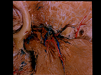
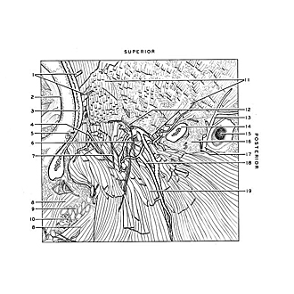
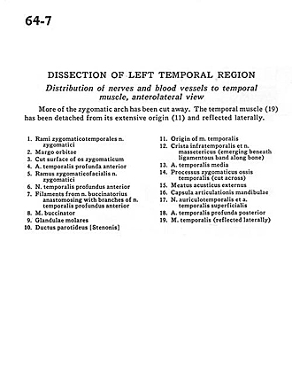
Dissection of left temporal region
Distribution of nerves and blood vessels to temporal muscle, anterolateral view
More of the zygomatic arch has been cut away. The temporal muscle (19) has been detached from its extensive origin (11) and reflected laterally.
- Zygomaticotemporal branches of zygomatic nerve
- Orbital margin
- Cut surface of zygomatic bone
- Deep anterior temporal artery
- Zygomaticofacial branch zygomatic nerve
- Anterior deep temporal nerve
- Filaments from buccinator nerve anastomosing with branches of anterior deep temporal nerve
- Buccinator muscle
- Molar glands
- Parotid duct
- Origin of temporalis muscle
- Infratemporal crest and masseteric nerve (emerging beneath ligamentous band along bone)
- Middle temporal artery
- Zygomatic process temporal bone (cut across)
- External acoustic meatus
- Joint capsule of mandible
- Auriculotemporal nerve and superficial temporal artery
- Deep posterior temporal artery
- Temporalis muscle (reflected laterally)


