
Bassett Collection of Stereoscopic Images of Human Anatomy
Dissection of left parotideomasseteric region
Deep relations of facial nerve
Image #63-4
KEYWORDS: Cheek, Exocrine and endocrine, Face, Peripheral nervous system.
Creative Commons
Stanford holds the copyright to the David L. Bassett anatomical images and has assigned Creative Commons license Attribution-Share Alike 4.0 International to all of the images.

For additional information regarding use and permissions, please contact the Medical History Center.

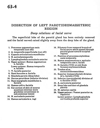
Dissection of left parotideomasseteric region
Deep relations of facial nerve
The superficial lobe of the parotid gland has been entirely removed and the facial nerved raised slightly away from the deep hole of the gland.
- Zygomatic process temporal bone (cut off)
- Superficial temporal artery (cut off)
- Articular capsule of mandible
- Auriculotemporal nerve
- Anterior auricular lymph node
- Upper pointer: Zygomatic branches of facial nerve Lower pointer: Temporal branch facial nerve
- Posterior facial vein
- Buccal branches of facial nerve
- Parotid gland (deep lobe)
- Parotid duct (inferior division)
- Marginal mandibular branches facial nerve
- Superficial branch facial nerve
- Cut section of skin of external acoustic meatus (note ceruminous gland in subcutaneous tissue)
- Posterior auricular nerve
- Auricular branch vagus nerve
- Filament from temporal branch of facial nerve which passed through parotid gland toward occipital region)
- Position of styloid process temporal bone
- Anastomotic branch auriculotemporal nerve with facial nerve
- Facial nerve (VII) at exit from stylomastoid foramen
- Tendon of sternocleidomastoid muscle
- Superior (temporofacial) division of facial nerve (VII)
- Inferior (cervicofacial) division of facial nerve (VII)
- Posterior auricular artery
- Deep lobe and duct of parotid gland
- Splenius capitis muscle
- Upper pointer: Posterior belly of digastric muscle Lower pointer: Superior deep cervical lymph node