
Bassett Collection of Stereoscopic Images of Human Anatomy
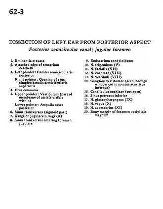
Dissection of left ear from posterior aspect
Posterior semicircular canal; jugular foramen
Image #62-3
KEYWORDS: Bones cartilage joints, Ear.
Creative Commons
Stanford holds the copyright to the David L. Bassett anatomical images and has assigned Creative Commons license Attribution-Share Alike 4.0 International to all of the images.

For additional information regarding use and permissions, please contact the Medical History Center.

Dissection of left ear from posterior aspect
Posterior semicircular canal; jugular foramen
- Arcuate eminence
- Attached edge of tentorium cerebelli
- Left pointer: Posterior semicircular canal Right pointer: Opening of crus simplex superior semicircular canal
- Common crus
- Upper pointer: Vestibule (part of membrane of utricle visible within) Lower pointer: Posterior osseous ampulla
- Transverse sinus (sigmoid part)
- Jugular ganglion of vagus nerve (X)
- Transverse sinus entering jugular foramen
- Condyloid emissary
- Trigeminal nerve (V)
- Facial nerve (VII)
- Vestibulocochlear nerve (VIII) (cochlear part)
- Vestibulocochlear nerve (VIII) (vestibular part)
- Vestibular ganglion (seen through window cut in internal acoustic meatus)
- Cochlear canaliculus (cut open)
- Inferior petrosal sinus
- Glossopharyngeal nerve (IX)
- Vagus nerve (X)
- Accessory nerve (XI)
- Bony margin of foramen magnum