
Bassett Collection of Stereoscopic Images of Human Anatomy
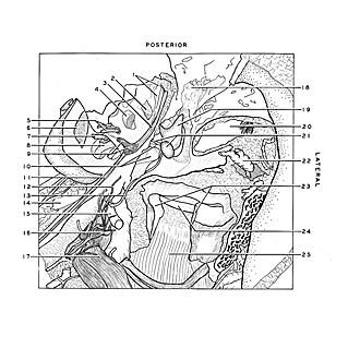
Dissection of left ear from superior aspect
Relations of tympanic cavity, facial canal and vestibule
Image #61-6
KEYWORDS: Bones cartilage joints, Ear.
Creative Commons
Stanford holds the copyright to the David L. Bassett anatomical images and has assigned Creative Commons license Attribution-Share Alike 4.0 International to all of the images.

For additional information regarding use and permissions, please contact the Medical History Center.
Dissection of left ear from superior aspect
Relations of tympanic cavity, facial canal and vestibule
The lateral semicircular canal has been cut away and the vestibule widely opened. The facial canal has been opened and it course above the posterior part of the tympanic cavity shown.
- Stapedius nerve and muscle
- Tympanic sinus
- Facial nerve (VII) (within facial canal)
- Vestibule
- Vestibulocochlear nerve (VIII) (vestibular part) (superior and inferior branches)
- Internal acoustic meatus
- Vestibulocochlear nerve (VIII) (cochlear part)
- Modiolus
- Upper pointer: Central end of facial nerve (Vll) (cut off) Lower pointer: Geniculate ganglion
- Upper pointer: Tendon of tensor tympani muscle Lower pointer: Tympanic membrane
- Major superficial petrosal nerve
- Minor superficial petrosal nerve
- Chorda tympani
- Internal carotid artery
- Middle meningeal artery
- Otic ganglion
- Mandibular nerve (V)
- Tympanic antrum
- Short crus of incus
- External acoustic meatus (note transition from thick to thin skin at junction of cartilaginous and bony parts of wall of meatus)
- Upper pointer: Manubrium of malleus Lower pointer: Capitulum of malleus
- Veins of external auditory meatus tributary to posterior facial vein
- Articular disc
- Capitulum (condylar process) of mandible
- External pterygoid muscle