Bassett Collection of Stereoscopic Images of Human Anatomy
Dissection of left ear from superior aspect
Tympanic cavity and labyrinth, superolateral view
Image #61-3
KEYWORDS: Bones cartilage joints, Ear.
Creative Commons
Stanford holds the copyright to the David L. Bassett anatomical images and has assigned Creative Commons license Attribution-Share Alike 4.0 International to all of the images.

For additional information regarding use and permissions, please contact the Medical History Center.

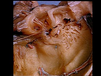
Dissection of left ear from superior aspect
Tympanic cavity and labyrinth, superolateral view
- Dissected area on posterior aspect of petrous part of temporal bone
- Common crus
- Upper pointer: Subarcuate fossa Lower pointer: Vestibule (membrane of utricle visible)
- Facial nerve (VII)
- Upper (utriculoampullar) division of vestibulocochlear nerve (VIII)
- Cochlea (pointer on modiolus)
- Geniculate ganglion
- Greater superficial petrosal nerve
- Upper pointer: Tendon of tensor tympani muscle Lower pointer: Position of anterior process of malleus
- Lessor superficial petrosal nerve
- Communicating branch between above nerve (10) and plexus along middle meningeal artery
- Middle meningeal artery (cut off)
- Semilunar ganglion (reflected laterally)
- Lateral semicircular canal
- Superior semicircular canal
- Tympanic antrum
- Posterior ligament of incus
- Upper pointer: Long crus of incus Lower pointer: Body incus
- Fold of incus
- Capitulum of malleus
- Lateral margin of tympanic cavity
- Mastoid cells