
Bassett Collection of Stereoscopic Images of Human Anatomy
Dissection of left ear from superior aspect
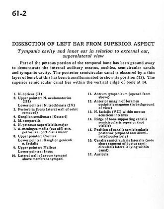
Tympanic cavity and inner ear in relation to external ear, superolateral view
Image #61-2
KEYWORDS: Bones cartilage joints, Ear.
Creative Commons
Stanford holds the copyright to the David L. Bassett anatomical images and has assigned Creative Commons license Attribution-Share Alike 4.0 International to all of the images.

For additional information regarding use and permissions, please contact the Medical History Center.

Dissection of left ear from superior aspect
Tympanic cavity and inner ear in relation to external ear, superolateral view
Part of the petrous portion of the temporal bone has been ground away to demonstrate the internal auditory meatus, cochlea, semicircular canals and tympanic cavity. The posterior semicircular canal is obscured by a thin layer of bone but this has been transilluminated to show its position (15). The superior semicircular canal lies within the vertical edge of bone at 14.
- Optic nerve (II)
- Upper pointer: Oculomotor nerve (III) Lower pointer: Trochlear nerve (IV)
- Periorbita (bony lateral wall of orbit removed)
- Semilunar ganglion (trigeminal)
- Temporalis muscle
- Greater superficial petrosal nerve
- Middle meningeal artery (cut off) and Lessor superficial petrosal nerve
- Upper pointer: Cochlea Lower pointer: Geniculate ganglion facial nerve
- Upper pointer: Malleus Lower pointer: Incus
- Lateral wall of tympanic cavity above tympanic membrane
- Tympanic antrum (opened from above)
- Anterior margin of foramen magnum (in background of view)
- Facial nerve (VII) within internal acoustic meatus
- Ridge of bone supporting superior semicircular canal (not visible)
- Position of posterior semicircular canal (exposed and illuminated posteriorly)
- Lateral semicircular canal (note short segment of lateral semicircular duct lying within canal)
- Auricle