Bassett Collection of Stereoscopic Images of Human Anatomy
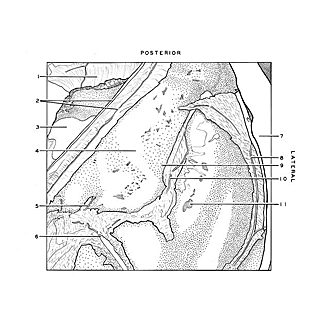
Dissection of left ear from superior aspect
Anterior surface of petrous part of temporal bone
Image #61-1
KEYWORDS: Bones cartilage joints, Ear.
Creative Commons
Stanford holds the copyright to the David L. Bassett anatomical images and has assigned Creative Commons license Attribution-Share Alike 4.0 International to all of the images.
For additional information regarding use and permissions, please contact the Medical History Center.
Dissection of left ear from superior aspect
Anterior surface of petrous part of temporal bone
The dura mater has been removed except in areas adjacent to blood vessels. Dark patches visible in the bone are caused by underlying air cells (11).
- Sigmoid part of transverse sinus
- Cut edges of tentorium enclosing superior petrosal sinus
- Area from which bone has been cut away
- Arcuate eminence
- Position of hiatus facial canal (note branch of middle meningeal artery entering hiatus)
- Middle meningeal artery
- Cut edge of calvaria
- Posterior branch of middle meningeal artery
- Tegmen tympani
- Position of Petrosquamous fissure
- Mastoid cell (visible beneath surface of bone)


