Bassett Collection of Stereoscopic Images of Human Anatomy
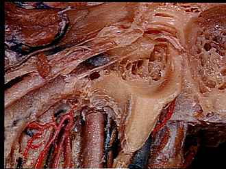
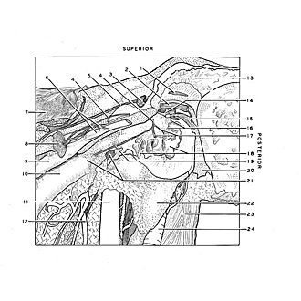
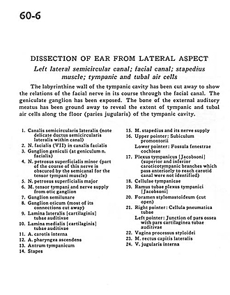
Dissection of ear from lateral aspect
Left lateral semicircular canal; facial canal; stapedius muscle; tympanic and tubal air cells
Image #60-6
KEYWORDS: Bones cartilage joints, Ear, Peripheral nervous system.
Creative Commons
Stanford holds the copyright to the David L. Bassett anatomical images and has assigned Creative Commons license Attribution-Share Alike 4.0 International to all of the images.

For additional information regarding use and permissions, please contact the Medical History Center.
Dissection of ear from lateral aspect
Left lateral semicircular canal; facial canal; stapedius muscle; tympanic and tubal air cells
The labyrinthine wall of the tympanic cavity has been cut away to show the relations of the facial nerve in its course through the facial canal. The geniculate ganglion has been exposed. The bone of the external auditory meatus has been ground away to reveal the extent of tympanic and tubal air cells along the floor (paries jugularis) of the tympanic cavity.
- Semicircular canal (lateral) (note delicate lateral semicircular duct within canal)
- Facial nerve (VII) in facial canal
- Geniculate ganglion (at geniculum facial nerve)
- Lessor superficial petrosal nerve (part of the course of this nerve is obscured by the semicanal for the tensor tympani muscle)
- Major superficial petrosal nerve
- Tensor tympani muscle and nerve supply from otic ganglion
- Semilunar ganglion
- Otic ganglion (most of its connections cut away)
- Lateral plate (cartilaginous) auditory tube
- Medial plate auditory tube
- Internal carotid artery
- Ascending pharyngeal artery
- Tympanic antrum
- Stapes
- Stapedius muscle and its nerve supply
- Upper pointer: Subiculum of promontory Lower pointer: Fenestrated cochlear fossa
- Tympanic plexus (superior and inferior caroticotympanic branches which pass anteriorly to reach carotid canal were not identified)
- Tympanic cells
- Tubal branches tympanic plexus
- Stylomastoid foramen (cut open)
- Right pointer: Tubal air cells Left pointer: Junction of bony part with cartilaginous part auditory tube
- Sheath for styloid process
- Rectus capitis lateralis muscle
- Internal jugular vein