
Bassett Collection of Stereoscopic Images of Human Anatomy
Dissection of ear from lateral aspect
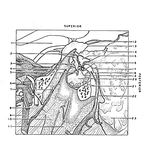
Left tympanic cavity, lateral view
Image #60-4
KEYWORDS: Bones cartilage joints, Ear, Peripheral nervous system.
Creative Commons
Stanford holds the copyright to the David L. Bassett anatomical images and has assigned Creative Commons license Attribution-Share Alike 4.0 International to all of the images.

For additional information regarding use and permissions, please contact the Medical History Center.

Dissection of ear from lateral aspect
Left tympanic cavity, lateral view
The tympanic membrane has been removed and more of the mastoid and petrous of the temporal bone resected. The facial nerve (17) and chorda tympani (7,16,18) have been exposed. The continuity of the tympanic antrum (14) with the mastoid air cells is visible.
- Base of peduncle (in background)
- Upper pointer: Superior ligament of malleus Lower pointer: Capitulum of malleus
- Upper pointer: Process of anterior malleus (covered by anterior malleolar plica) Lower pointer: Lateral process malleus (the superior recess, Prussak's space, is located just above this process and is closed laterally by a thin downward extension of squamous bone known as the scutum)
- Petrotympanic fissure
- Manubrium of malleus
- Upper pointer: Mandibular nerve (V) Lower pointer: Middle meningeal artery
- Chorda tympani (passing inferiorly to join lingual nerve)
- Tensor veli palatini muscle
- Lingual nerve
- Alar fascia
- Styloglossus muscle
- Arcuate eminence
- Epitympanic recess
- Tympanic antrum
- Left pointer: Body incus Right pointer: Posterior ligament of incus
- Left pointer: Chorda tympani within tympanic cavity Right pointer: Tendon of stapedius muscle
- Facial nerve (VII) within facial canal
- Chorda tympani within canaliculus
- Left pointer: Anterior crus of stapes Right pointer: Tympanic sinus
- Upper pointer: Fenestrated cochlear fossa (separated from tympanic sinus above by promontory) Lower pointer: Promontory (tympanic plexus visible beneath mucosa)
- Tympanic cells of jugular wall of tympanic cavity
- Position of stylomastoid foramen (opened)
- Mastoid process (dissected)