
Bassett Collection of Stereoscopic Images of Human Anatomy
Exploration of the brain from its basal aspect
Mammillothalamic tract; medial forebrain bundle; substantia nigra
Image #6-7
KEYWORDS: Brain, Diencephalon, Midbrain.
Creative Commons
Stanford holds the copyright to the David L. Bassett anatomical images and has assigned Creative Commons license Attribution-Share Alike 4.0 International to all of the images.

For additional information regarding use and permissions, please contact the Medical History Center.
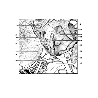
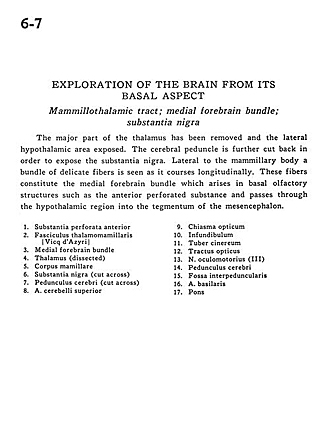
Exploration of the brain from its basal aspect
Mammillothalamic tract; medial forebrain bundle; substantia nigra
The major part of the thalamus has been removed and the lateral hypothalamic area exposed. The cerebral peduncle is further cut back in order to expose the substantia nigra. Lateral to the mammillary body a bundle of delicate fibers is seen as it courses longitudinally. These fibers constitute the medial forebrain bundle which arises in basal olfactory structures such as the anterior perforated substance and passes through the hypothalamic region into the tegmentum of the mesencephalon.
- Anterior perforated substance
- Mamillothalamic tract (Vicq d'Azyr)
- Medial forebrain bundle
- Thalamus (dissected)
- Mamillary body
- Substantia nigra (cut across)
- Cerebral peduncle (cut across)
- Superior cerebellar artery
- Optic chiasm
- Infundibulum
- Tuber cinereum
- Optic tract
- Oculomotor nerve (III)
- Cerebral peduncle
- Interpeduncular fossa
- Basilar artery
- Pons