Bassett Collection of Stereoscopic Images of Human Anatomy
Exploration of the brain from its basal aspect
Caudate nucleus and lateral border of thalamus
Image #6-4
KEYWORDS: Brain, Diencephalon, Telencephalon.
Creative Commons
Stanford holds the copyright to the David L. Bassett anatomical images and has assigned Creative Commons license Attribution-Share Alike 4.0 International to all of the images.

For additional information regarding use and permissions, please contact the Medical History Center.
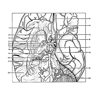
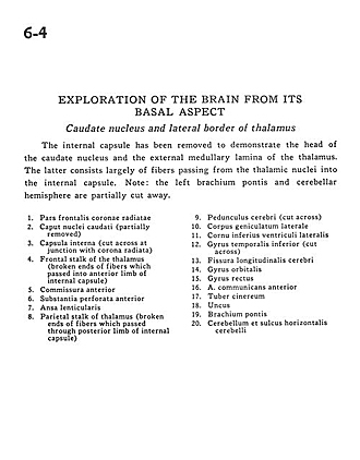
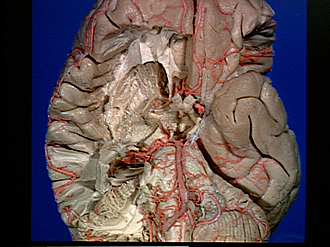
Exploration of the brain from its basal aspect
Caudate nucleus and lateral border of thalamus
The internal capsule has been removed to demonstrate the head of the caudate nucleus and the external medullary lamina of the thalamus. The latter consists largely of fibers passing from the thalamic nuclei into the internal capsule. Note
- Frontal part corona radiata
- Head of caudate nucleus (partially removed)
- Internal capsule (cut across at junction with corona radiata)
- Frontal stalk of the thalamus (broken ends of fibers which passed into anterior limb of internal capsule)
- Anterior commissure
- Anterior perforated substance
- Ansa lenticularis
- Parietal stalk of thalamus (broken ends of fibers which passed through posterior limb of internal capsule)
- Cerebral peduncle (cut across)
- Lateral geniculate body
- Inferior horn of lateral ventricle
- Inferior temporal gyrus (cut across)
- Longitudinal, fissure (cerebral)
- Orbital gyrus
- Straight gyrus
- Anterior communicating artery
- Tuber cinereum
- Uncus
- Brachium pontis (middle cerebellar peduncle)
- Cerebellum and horizontal cerebellar sulcus