Bassett Collection of Stereoscopic Images of Human Anatomy
Dissection of ear from lateral aspect
Right auricular cartilage, medial surface
Image #59-6
KEYWORDS: Bones cartilage joints, Ear.
Creative Commons
Stanford holds the copyright to the David L. Bassett anatomical images and has assigned Creative Commons license Attribution-Share Alike 4.0 International to all of the images.

For additional information regarding use and permissions, please contact the Medical History Center.
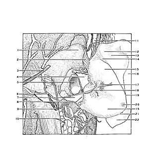

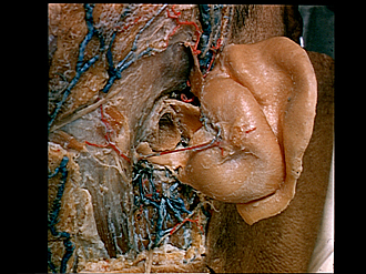
Dissection of ear from lateral aspect
Right auricular cartilage, medial surface
The cartilage has been retracted anterolaterally after removal of its perichondrium.
- Temporal fascia
- Superior auricular muscle
- Lesser occipital nerve
- Tragal plate
- External acoustic meatus
- Occipital branch posterior auricular artery
- Cartilaginous acoustic meatus
- Terminal incisure (pointer on cartilaginous isthmus)
- Posterior auricular nerve
- Parotid gland
- Superficial temporal vein and auriculotemporal nerve
- Eminence of triangular fossa
- Sulcus of upper crus of anthelix
- Transverse sulcus of anthelix
- Conchal eminence
- Scaphoid eminence
- Posterior auricular muscle and posterior auricular ligament (cut off)
- Posterior auricular branch auricular artery
- Fossa of anthelix
- Transverse auricular muscle
- Antitragicohelical fissure
- Tail of helix