Bassett Collection of Stereoscopic Images of Human Anatomy
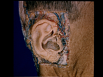
Dissection of ear from lateral aspect
Superficial structures of right auricle, lateral view
Image #59-3
KEYWORDS: Ear, Peripheral nervous system, Vasculature.
Creative Commons
Stanford holds the copyright to the David L. Bassett anatomical images and has assigned Creative Commons license Attribution-Share Alike 4.0 International to all of the images.

For additional information regarding use and permissions, please contact the Medical History Center.
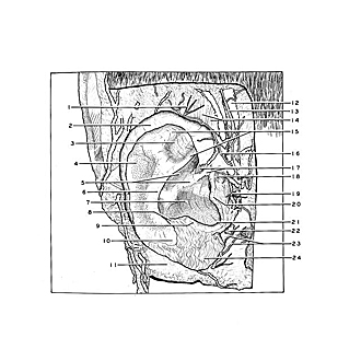

Dissection of ear from lateral aspect
Superficial structures of right auricle, lateral view
The skin has been removed from the auricle to demonstrate the subcutaneous vessels and nerves. The perichondrium remains intact.
- Lesser occipital nerve
- Helix
- Triangular fossa
- Scaphoid fossa
- Upper pointer: Cymba conchae Lower pointer: Crus of helix
- Anthelix
- Cavum conchae
- Posterior auricular sulcus
- Antitragus
- Tail of helix
- Superficial fascia covering sternocleidomastoid muscle
- Superficial temporal vein
- Auriculotemporal nerve
- Superior auricular muscle
- Crura of anthelix
- Anterior auricular branch of superficial temporal artery
- Upper pointer: Helicis minor muscle Lower pointer: Anterior incisura
- Tragicus muscle
- Anterior auricular nerves
- Tragus
- Intertragic incisure
- Anterior auricular lymph nodes
- Greater auricular nerve (anterior branch)
- Lobule