Bassett Collection of Stereoscopic Images of Human Anatomy
Microradiograph of eye; central optic pathways and related structures
Relations of optic pathways at base of brain.
Image #58A-7
KEYWORDS: Brain, Diencephalon, Eye, Face, Peripheral nervous system, Telencephalon, Temporal lobe, Vasculature.
Creative Commons
Stanford holds the copyright to the David L. Bassett anatomical images and has assigned Creative Commons license Attribution-Share Alike 4.0 International to all of the images.

For additional information regarding use and permissions, please contact the Medical History Center.
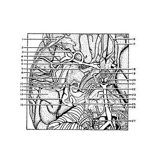

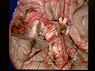
Microradiograph of eye; central optic pathways and related structures
Relations of optic pathways at base of brain.
In this specimen, selected from a series of brain dissections included in Section I of the Atlas, the brain is photographed from its inferior aspect. The anterior part of the right temporal lobe has been removed so that the middle cerebral artery (6), the anterior choroidal artery (18), the inferior horn of the lateral ventricle (10), and the optic tract (19) life exposed.
- Meninges related to temporal pole
- Orbitofrontal branch of middle cerebral artery
- Anterior temporal artery
- Insula
- Frontal part of operculum
- Medial cerebral artery
- Striate artery
- Artery of precentral sulcus
- Inferior occipitofrontal fasciculus (cut obliquely)
- Inferior horn of lateral ventricle
- Tapetum
- Medial temporal gyrus
- Inferior temporal gyrus
- Inferior temporal sulcus
- Fusiform gyrus
- Orbital branch of anterior cerebral artery
- Olfactory tract
- Anterior choroid artery
- Optic tract
- Mamillary body
- Cerebral peduncle
- Hippocampus (cut across)
- Pons
- Trigeminal nerve (V)
- Collateral fissure
- Basilar artery
- Olive
- [Legend above restored translation from Latin]