Bassett Collection of Stereoscopic Images of Human Anatomy
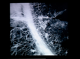
Microradiograph of eye; central optic pathways and related structures
Microradiography of limbic region of eye, viewed from in front
Image #58A-1
KEYWORDS: Eye, Face, Vasculature.
Creative Commons
Stanford holds the copyright to the David L. Bassett anatomical images and has assigned Creative Commons license Attribution-Share Alike 4.0 International to all of the images.
For additional information regarding use and permissions, please contact the Medical History Center.
Microradiograph of eye; central optic pathways and related structures
Microradiography of limbic region of eye, viewed from in front
This micrograph was obtained through the courtesy of Dr. H.H. Pattee and Dr. L.K. Garron who prepared and radiographed the specimen. Reference to be made to their article entitled "Stereomicroradiography of the limbal region of human eye" in X-Ray Microscopy and Microradiography, Academic Press, 1957. Thorotrast was injected into the canal of Schlemm (1) under low pressure. The thorium filled the canal and passed into the vascular network of the sclera (2) and the superficial vascular network of the corne al margin. The specimen was frozen in liquid nitrogen and lyophilized prior to radiography.
- Canal of Schlemm
- Sclera
- Cornea
- Superficial vascular network of cornea


