Bassett Collection of Stereoscopic Images of Human Anatomy
Collection Home
- Abdomen See All
- Overview 33
- Adrenal Gland 29
- Bones Cartilage Joints 4
- Central Nervous System 2
- Fascia 16
- Gallbladder 27
- Kidney 29
- Large Intestine 14
- Liver 29
- Lymphatics 12
- Muscles & Tendons 55
- Pancreas 27
- Peripheral Nervous System 56
- Small Intestine 11
- Spleen 29
- Stomach 15
- Vasculature 70
- Back See All
- Overview 5
- Bones Joints Cartilage 34
- Central Nervous System 22
- Cervical Region 42
- Lumbar Region 52
- Muscles & Tendons 40
- Meninges 5
- Peripheral Nervous System 6
- Sacral Region 37
- Thoracic Region 55
- Vasculature 17
- Vertebral Column 107
- Head See All
- Overview 49
- Bones Cartilage Joints 176
- Brain 225
- Cerebellum 51
- Cheek 41
- Connective Tissue 49
- Diencephalon 71
- Ear 37
- Exocrine & Endocrine 15
- Eye 62
- Face 153
- Frontal Lobe 22
- Medulla 32
- Meninges 23
- Midbrain 54
- Mouth 63
- Muscles & Tendons 70
- Nose 20
- Occipital Lobe 21
- Parietal Lobe 20
- Peripheral Nervous System 126
- Pons 37
- Scalp 16
- Telencephalon 134
- Temporal Lobe 28
- Vasculature 131
- Ventricules 61
- Female Pelvis See All
- Overview 1
- Anal Canal 6
- Bones Joints Cartilage 48
- Central Nervous System 26
- External Genitalia 9
- Large Intestine 6
- Muscles& Tendons 57
- Ovary 18
- Perineum 11
- Peripheral Nervous System 20
- Urinary Tract 14
- Uterus 16
- Vagina 14
- Vasculature 40
- Lower Extremity See All
- Ankle 42
- Bones Joints Cartilage 75
- Fascia 19
- Foot & Toes 78
- Knee 22
- Leg 41
- Muscles & Tendons 152
- Peripheral Nervous System 80
- Thigh 58
- Vasculature 53
- Male Pelvis See All
- Anal Canal 2
- Bones Joints Cartilage 33
- Central Nervous System 10
- Large Intestine 2
- Muscles & Tendons 37
- Perineum 3
- Peripheral Nervous System 14
- Prostate 2
- Urinary Tract 6
- Vasculature 33
- Neck See All
- Overview 13
- Bones Cartilage Joints 35
- Central Nervous System 9
- Cervical Vertebrae 23
- Esophagus 5
- Exocrine & Endocrine 20
- Fascia & Connective Tissue 37
- Lymphatics 9
- Meninges 5
- Muscles & Tendons 54
- Peripheral Nervous System 55
- Pharynx 17
- Throat 51
- Trachea 2
- Vasculature 47
- Pelvis See All
- Overview 2
- Anal Canal 6
- Bones Joints Cartilage 44
- Central Nervous System 27
- External Genitalia 10
- Female 1
- Large Intestine 6
- Muscles & Tendons 57
- Ovary 18
- Perineum 10
- Peripheral Nervous System 20
- Urinary Tract 14
- Uterus 16
- Vagina 14
- Vasculature 46
- Thorax See All
- Overview 7
- Bones Joints Cartilage 33
- Breast 7
- Central Nervous System 6
- Diaphragm 8
- Esophagus 10
- Fascia & Connective Tissue 12
- Heart 46
- Left Heart 33
- Left Lung 21
- Lung 39
- Lymphatics 9
- Mediastinum 23
- Muscles & Tendons 28
- Pericardial Sac 25
- Peripheral Nervous System 32
- Pleura 11
- Rib Cage 16
- Right Heart 31
- Right Lung 17
- Skin 2
- Thymus 2
- Vasculature 43
- Vertebral Column 20
Creative Commons
Stanford holds the copyright to the David L. Bassett anatomical images and has assigned Creative Commons license Attribution-Share Alike 4.0 International to all of the images.

For additional information regarding use and permissions, please contact the Medical History Center.
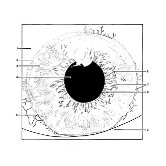
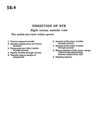
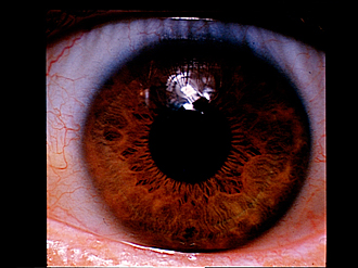
Dissection of eye
Right cornea, anterior view
The eyelids have been widely opened.
- Tunica bulbar conjunctiva
- Annulus conjunctiva (at limbus of cornea)
- Anterior surface of iris (visible through cornea)
- Pupil (visible through cornea)
- Vascular loop at margin of conjunctiva
- Annulus iris minor (visible through cornea)
- Annulus iris major (visible through cornea)
- Pupillary-iris margin (note: ciliary margin of iris obscured by annulus conjunctiva (2))
- Inferior palpebra