Bassett Collection of Stereoscopic Images of Human Anatomy
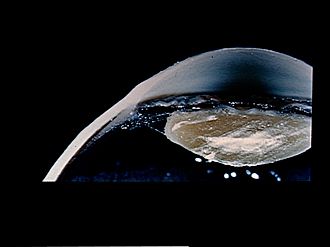
Dissection of eye
Ciliary body, ciliary zonule and lens of the left eye, horizontal section
Image #58-3
KEYWORDS: Eye, Face.
Creative Commons
Stanford holds the copyright to the David L. Bassett anatomical images and has assigned Creative Commons license Attribution-Share Alike 4.0 International to all of the images.

For additional information regarding use and permissions, please contact the Medical History Center.
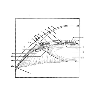
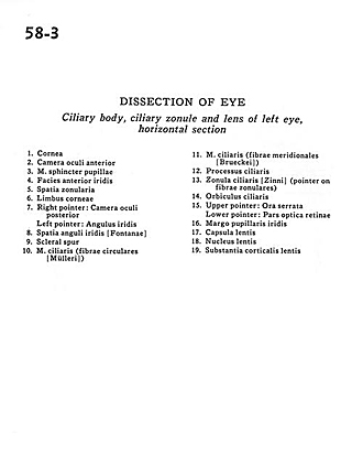
Dissection of eye
Ciliary body, ciliary zonule and lens of the left eye, horizontal section
- Cornea
- Anterior chamber of eye
- Pupillary sphincter muscle
- Anterior surface of iris
- Zonular space
- Limbus of cornea
- Right pointer: Posterior chamber of eye Left pointer: Angle of iris
- Angular space of iris
- Scleral spur
- Ciliary muscle (circular fibers)
- Ciliary muscle (circular fibers)
- Ciliary process
- Zonula ciliaris (pointer on zonular fibers)
- Orbiculus ciliaris
- Upper pointer: Ora serrata Lower pointer: Optic (visual) part of retina
- Iris-pupillary margin
- Capsule of lens
- Nucleus of lens
- Substantia corticalis of lens