Bassett Collection of Stereoscopic Images of Human Anatomy
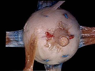
Insertion of extraocular muscles
Left eye, posterior view
Image #57-6
KEYWORDS: Connective tissue, Eye, Face, Muscles and tendons.
Creative Commons
Stanford holds the copyright to the David L. Bassett anatomical images and has assigned Creative Commons license Attribution-Share Alike 4.0 International to all of the images.

For additional information regarding use and permissions, please contact the Medical History Center.

Insertion of extraocular muscles
Left eye, posterior view
The fascia has been removed from the muscles and the eye, and the muscles retracted and stretched.
- Tendon of superior rectus muscle
- Sclera
- Sheath of optic nerve
- Upper pointer: Posterior ciliary artery (long) Lower pointer: Position of macula lutea of retina (marked by *)
- Tendon of lateral rectus muscle
- Short ciliary nerves
- Inferior oblique muscle
- Tendon of superior oblique muscle
- Superior vorticose veins
- Posterior ciliary arteries (short)
- Medial rectus muscle
- Optic nerve (II)
- Inferior vorticose veins
- Tendon of Inferior rectus muscle