Bassett Collection of Stereoscopic Images of Human Anatomy
Dissection of left orbit from a lateral approach
Eye and sheath of optic nerve; ophthalmic artery and ciliary ganglion
Image #56-4
KEYWORDS: Connective tissue, Eye, Face, Muscles and tendons, Peripheral nervous system, Vasculature.
Creative Commons
Stanford holds the copyright to the David L. Bassett anatomical images and has assigned Creative Commons license Attribution-Share Alike 4.0 International to all of the images.
For additional information regarding use and permissions, please contact the Medical History Center.
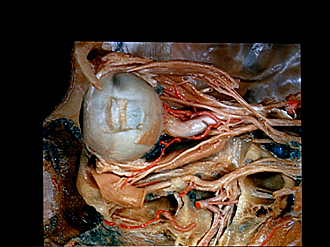
Dissection of left orbit from a lateral approach
Eye and sheath of optic nerve; ophthalmic artery and ciliary ganglion
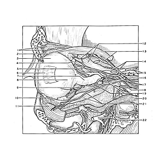
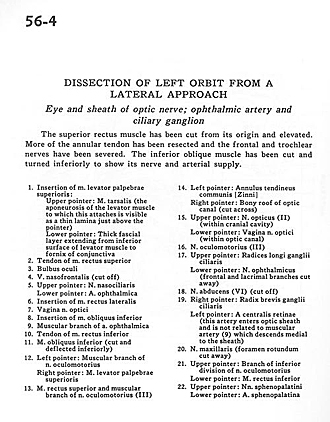
The superior rectus muscle has been cut from its origin and elevated. More of the annular tendon has been resected and the frontal and trochlear nerves have been severed. The inferior oblique muscle has been cut and turned inferiorly to show its nerve and arterial supply.
- Insertion of levator palpebrae superioris muscle: Upper pointer: Tarsalis muscle (the aponeurosis of the levator muscle to which this attaches is visible as a thin lamina just above the pointer) Lower pointer: Thick fascial layer extending from inferior surface of levator muscle to fornix of conjunctiva
- Tendon of superior rectus muscle
- Eyeball
- Nasofrontal vein (cut off)
- Upper pointer: Nasociliary nerve Lower pointer: Ophthalmic artery
- Insertion of lateral rectus muscle
- Sheath of optic nerve
- Insertion of inferior oblique muscle
- Muscular branch of ophthalmic artery
- Tendon of inferior rectus muscle
- Inferior oblique muscle (cut and deflected inferiorly)
- Left pointer: Muscular branch of Oculomotor nerve Right pointer: Levator palpebrae superioris muscle
- Superior rectus muscle and muscular branch of oculomotor nerve (III)
- Left pointer: Common annular tendon Right pointer: Bony roof of optic canal (cut across)
- Upper pointer: Optic nerve (II) (within cranial cavity) Lower pointer: Sheath of optic nerve (within optic canal)
- Oculomotor nerve (III)
- Upper pointer: Long roots of diary ganglion Lower pointer: Ophthalmic nerve (frontal and lacrimal branches cut away)
- Abducens nerve (VI) (cut off)
- Right pointer: Short branches of ciliary ganglion Left pointer: Central retinal artery (this artery enters optic sheath and is not related to muscular artery (9) which descends medial to the sheath)
- Maxillary nerve (foramen rotundum cut away)
- Upper pointer: Branch of inferior division of oculomotor nerve Lower pointer: Inferior rectus muscle
- Upper pointer: Sphenopalatine nerves Lower pointer: Sphenopalatine artery


