Bassett Collection of Stereoscopic Images of Human Anatomy
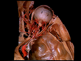
Dissection of right orbit from a superior approach
Branches of ophthalmic artery
Image #54-6
KEYWORDS: Connective tissue, Eye, Face, Peripheral nervous system, Vasculature.
Creative Commons
Stanford holds the copyright to the David L. Bassett anatomical images and has assigned Creative Commons license Attribution-Share Alike 4.0 International to all of the images.

For additional information regarding use and permissions, please contact the Medical History Center.


Dissection of right orbit from a superior approach
Branches of ophthalmic artery
The optic nerve has been cut and the eye turned anteriorly. The central end of the optic nerve has been displaced from the optic canal to illustrate the course of the ophthalmic artery in a separate dural investment inferior to the nerve. Branches of this artery within the orbit presented such a complex pattern that it was necessary to displace the artery medially from its original position to demonstrate their arrangement. Branches to structures in the superior part of the orbit have been cut off. In the accompanying drawing smaller arterial branches have been omitted for the sake of simplicity.
- Frontal artery
- Medial check ligament
- Posterior ciliary artery (long) (within sclera)
- Anterior ethmoidal artery
- Medial rectus muscle
- Posterior ciliary artery
- Inferior muscular branch of ophthalmic artery (branches pass to medial, inferior and lateral rectus muscles and inferior oblique muscle)
- Posterior ethmoidal artery
- Lacrimal artery (cut off)
- Ciliary ganglion
- Optic nerve (II) (elevated
- Ophthalmic artery entering optic canal
- Internal carotid artery
- Insertion of superior oblique muscle
- Lacrimal gland (inferior)
- Lateral check ligament
- Posterior ciliary arteries (short)
- Optic nerve (ll) within sheath
- Lateral rectus muscle
- Central artery of retina (continuity of artery interrupted in drawing but not in specimen)
- Posterior ciliary artery
- Origin of superior rectus muscle (cut off)
- Nerves entering orbit through superior orbital fissure