Bassett Collection of Stereoscopic Images of Human Anatomy
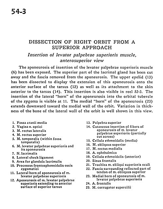
Dissection of right orbit from a superior approach
Insertion of levator palpebrae superioris muscle, anterosuperior view
Image #54-3
KEYWORDS: Eye, Face, Muscles and tendons.
Creative Commons
Stanford holds the copyright to the David L. Bassett anatomical images and has assigned Creative Commons license Attribution-Share Alike 4.0 International to all of the images.

For additional information regarding use and permissions, please contact the Medical History Center.
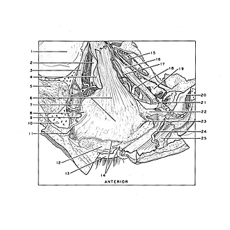
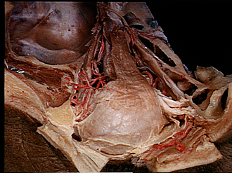
Dissection of right orbit from a superior approach
Insertion of levator palpebrae superioris muscle, anterosuperior view
The aponeurosis of insertion of the levator palpebrae superioris muscle (6) has been exposed. The superior part of the lacrimal gland has been cut away and the fascia removed from the aponeurosis. The upper eyelid (13) has been dissected to display the extension of this aponeurosis onto the anterior surface of the tarsus (12) as well as its attachment to the skin anterior to the tarsus (14). This insertion is also visible in reel 52-3. The insertion of the lateral "horn" of the aponeurosis into the orbital tubercle of the zygoma is visible at 11. The medial "horn" of the aponeurosis (23) extends downward toward the medial wall of the orbit. Variation in thickness of the bone of the lateral wall of the orbit is well shown in this view.
- Middle cranial fossa
- Sheath of optic nerve
- Lateral rectus muscle
- Superior rectus muscle
- Temporalis muscle (within temporal fossa)
- Levator palpebrae superioris muscle and its aponeurosis
- Lacrimal nerve
- Lateral check ligament
- Area for lacrimal gland
- Frontosphenoidal process of zygomatic bone
- Lateral horn of aponeurosis of levator palpebrae superioris muscle
- Aponeurosis of levator palpebrae superioris muscle extending to anterior surface of superior tarsus
- Superior palpebra
- Cutaneous insertion of fibers of aponeurosis of levator palpebrae superioris muscle (partially cut across)
- Ethmoidal cell (medial)
- Superior oblique muscle
- Medial rectus muscle
- Ophthalmic artery
- Ethmoidal cell (anterior)
- Frontal sinus
- Superior oblique muscle
- Fascia surrounding reflected part of tendon of superior oblique muscle
- Medial horn of aponeurosis of Ievator palpebrae superioris muscle
- Frontal artery
- Corrugator supercilii muscle