
Bassett Collection of Stereoscopic Images of Human Anatomy
Dissection of left orbit from an anterior approach
Orbital septum; corrugator supercilii muscle
Image #52-4
KEYWORDS: Connective tissue, Eye, Face, Muscles and tendons, Peripheral nervous system, Vasculature.
Creative Commons
Stanford holds the copyright to the David L. Bassett anatomical images and has assigned Creative Commons license Attribution-Share Alike 4.0 International to all of the images.

For additional information regarding use and permissions, please contact the Medical History Center.
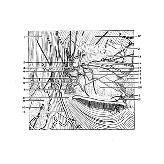
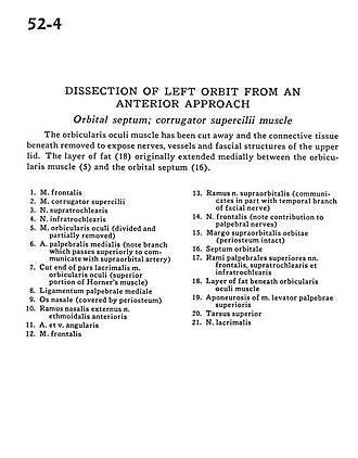
Dissection of left orbit from an anterior approach
Orbital septum; corrugator supercilii muscle
The orbicularis oculi muscle has been cut away and the connective tissue beneath removed to expose nerves, vessels and fascial structures of the upper lid. The layer of fat (18) originally extended medially between the orbicularis muscle (5) and the orbital septum (16).
- Frontalis muscle
- Corrugator supercilii muscle
- Supratrochlear nerve
- Infratrochlear nerve
- Orbicularis oculi muscle (divided and partially removed)
- Medial palpebral artery (note branch which passes superiorly to communicate with supraorbital artery)
- Cut end of lacrimal part orbicularis oculi muscle (superior portion of Horner's muscle)
- Medial palpebral ligament
- Nasal bone (covered by periosteum)
- External nasal branch anterior ethmoidal nerve
- Angular artery and vein
- Frontalis muscle
- Branch supraorbital nerve (communicates in part with temporal branch of facial nerve)
- Frontal nerve (note contribution to palpebral nerves)
- Supraorbital margin of orbit (periosteum intact)
- Orbital septum
- Superior palpebral branches of frontal, supratrochlear, and infratrochlear nerves
- Layer of fat beneath orbicularis oculi muscle
- Aponeurosis of levator palpebrae superioris muscle
- Superior tarsus
- Lacrimal nerve