Bassett Collection of Stereoscopic Images of Human Anatomy
Floor of cranial cavity
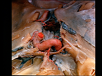
Middle cranial fossa; dissection of left cavernous sinus (continued); internal carotid artery; hypophysis sectioned in midsagittal plane
Image #51-3
KEYWORDS: Bones cartilage joints, Peripheral nervous system, Vasculature.
Creative Commons
Stanford holds the copyright to the David L. Bassett anatomical images and has assigned Creative Commons license Attribution-Share Alike 4.0 International to all of the images.

For additional information regarding use and permissions, please contact the Medical History Center.

Floor of cranial cavity
Middle cranial fossa; dissection of left cavernous sinus (continued); internal carotid artery; hypophysis sectioned in midsagittal plane
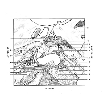
Communicating branches between the sympathetic plexus on the internal carotid artery and the abducens nerve have been exposed and the ophthalmic sympathetic nerve (2) traced onto the ophthalmic artery. The course of the internal carotid artery (13) is seen in relation to the sella turcica (12).
- Hypophyseal branch of internal carotid artery right
- Ophthalmic artery and ophthalmic nerve plexus
- Optic nerve (II) (anterior clinoid process cut away)
- Upper pointer: Oculomotor nerve (III) Lower pointer: Trochlear nerve (IV)
- Ophthalmic nerve entering superior orbital fissure
- Lacrimal nerve
- Superior ophthalmic vein (bone of lateral wall of orbit cut away)
- Posterior clinoid process
- Upper pointer: Infundibulum Lower pointer: Posterior pituitary (hypophysis)
- Anterior pituitary (hypophysis)
- Basilar venous plexus
- Sella turcica (dura mater stripped away posteriorly to expose small veins and artery)
- Internal carotid artery
- Abducens nerve (VI)
- Cavernous nerve plexus
- Branch of internal carotid artery to semilunar ganglion and meninges of cavernous sinus
- Sphenopetrosal ligament
- Semilunar ganglion