
Bassett Collection of Stereoscopic Images of Human Anatomy
Floor of cranial cavity
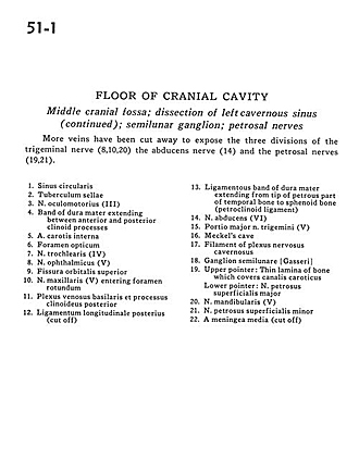
Middle cranial fossa; dissection of left cavernous sinus (continued); semilunar ganglion; petrosal nerves
Image #51-1
KEYWORDS: Bones cartilage joints, Peripheral nervous system, Vasculature.
Creative Commons
Stanford holds the copyright to the David L. Bassett anatomical images and has assigned Creative Commons license Attribution-Share Alike 4.0 International to all of the images.

For additional information regarding use and permissions, please contact the Medical History Center.

Floor of cranial cavity
Middle cranial fossa; dissection of left cavernous sinus (continued); semilunar ganglion; petrosal nerves
More veins have been cut away to expose the three divisions of the trigeminal nerve (8,10,20) the abducens nerve (14) and the petrosal nerves (19,21).
- Circular sinus
- Tuberculum sellae
- Oculomotor nerve (III)
- Band of dura mater extending between anterior and posterior clinoid processes
- Internal carotid artery
- Optic foramen
- Trochlear nerve (IV)
- Ophthalmic nerve (V1)
- Superior orbital fissure
- Maxillary nerve (V2) entering foramen rotundum
- Basilar venous plexus and posterior clinoid process
- Posterior longitudinal ligament (cut off)
- Ligamentous band of dura mater extending from tip of petrous part of temporal bone to sphenoid bone (petrodinoid ligament)
- Abducens nerve (VI)
- Major portion of trigeminal nerve (V)
- Meckel's cave
- Filament of cavernous nerve plexus
- Semilunar ganglion (trigeminal)
- Upper pointer: Thin lamina of bone which covers carotid canal Lower pointer: Major superficial petrous nerve
- Mandibular nerve (V3)
- Lesser superficial petrosal nerve
- Middle meningeal artery (cut off)