Bassett Collection of Stereoscopic Images of Human Anatomy
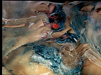
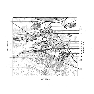
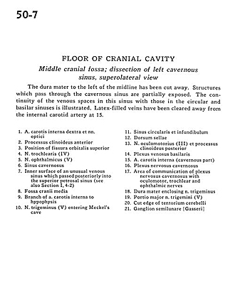
Floor of cranial cavity
Middle cranial fossa; dissection of left cavernous sinus, superolateral view
Image #50-7
KEYWORDS: Bones cartilage joints, Peripheral nervous system, Vasculature.
Creative Commons
Stanford holds the copyright to the David L. Bassett anatomical images and has assigned Creative Commons license Attribution-Share Alike 4.0 International to all of the images.

For additional information regarding use and permissions, please contact the Medical History Center.
Floor of cranial cavity
Middle cranial fossa; dissection of left cavernous sinus, superolateral view
The dura mater to the left of the midline has been cut away. Structures which pass through the cavernous sinus are partially exposed. The continuity of the venous spaces in this sinus with those in the circular and basilar sinuses is illustrated. Latex-filled veins have been cleared away from the internal carotid artery at 15.
- Internal carotid artery right and optic nerve
- Anterior clinoid process
- Position of superior orbital fissure
- Trochlear nerve (IV)
- Ophthalmic nerve (V)
- Cavernous sinus
- Inner surface of an unusual venous sinus which passed posteriorly into the superior petrosal sinus (see also Section I, 42)
- Middle cranial fossa
- Branch of internal carotid artery to hypophysis
- Trigeminal nerve (V) entering Meckel's cave
- Circular sinus and infundibulum
- Dorsum sellae
- Oculomotor nerve (III) and posterior clinoid process
- Basilar venous plexus
- Internal carotid artery (cavernous part)
- Cavernous nerve plexus
- Area of communication of cavernous nerve plexus with oculomotor, trochlear and ophthalmic nerves
- Dura mater enclosing trigeminal nerve
- Greater part of trigeminal nerve (V)
- Cut edge of tentorium cerebelli
- Semilunar ganglion (trigeminal)