Bassett Collection of Stereoscopic Images of Human Anatomy
Floor of cranial cavity
Structures inferior to anterior and middle cranial fossae; orbit and sinuses opened
Image #50-5
KEYWORDS: Bones cartilage joints, Peripheral nervous system, Vasculature.
Creative Commons
Stanford holds the copyright to the David L. Bassett anatomical images and has assigned Creative Commons license Attribution-Share Alike 4.0 International to all of the images.

For additional information regarding use and permissions, please contact the Medical History Center.


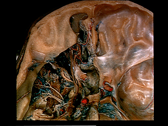
Floor of cranial cavity
Structures inferior to anterior and middle cranial fossae; orbit and sinuses opened
Views of the dissection of the contents of the orbit from this approach are found in 54-1 ff.
- Frontal sinus
- Infundibulum leading into nasofrontal duct
- Upper pointer: Frontal nerve Lower pointer: Levator palpebrae superioris
- Posterior ethmoidal nerve (usually a branch of the ophthalmic nerve in this case the fibers accompany the trochlear nerve)
- Posterior ethmoidal artery in posterior ethmoidal foramen
- Lacrimal nerve and superior ophthalmic vein
- Periorbita (area of fusion with common annular tendon)
- Maxillary nerve (V2)
- Trochlear nerve (IV)
- Oculomotor nerve (III)
- Abducens nerve (VI)
- Vidian nerve of pterygoid canal (below floor of sphenoid sinus)
- Semilunar ganglion (trigeminal)
- Mandibular nerve (V3)
- Cavernous nerve plexus
- Ethmoidal cell (anterior)
- Anterior ethmoidal nerve
- Cribriform plate ethmoid bone
- Anterior ethmoidal artery (in anterior ethmoidal foramen)
- Ethmoidal cell (posterior)
- Posterior ethmoidal vein
- Upper pointer: Aperture of sphenoid sinus Lower pointer: Sphenoid sinus
- Optic nerve (ll)
- Internal carotid artery
- Upper pointer: Circular sinus Lower pointer: Hypophysis (left half removed)
- Sella turcica
- Dorsum sellae
- Basilar venous plexus