Bassett Collection of Stereoscopic Images of Human Anatomy
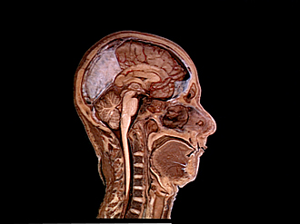
General orientation views of dissection
Midsagittal section of head and neck, right lateral view
Image #50-1
KEYWORDS: Bones cartilage joints, Brain, Cerebellum, Face, Frontal lobe, Medulla, Midbrain, Mouth, Muscles and tendons, Nose, Occipital lobe, Peripheral nervous system, Pons, Telencephalon, Vasculature, Central nervous system, Cervical vertebrae, Esophagus, Pharynx, Throat, Trachea, Overview.
Creative Commons
Stanford holds the copyright to the David L. Bassett anatomical images and has assigned Creative Commons license Attribution-Share Alike 4.0 International to all of the images.

For additional information regarding use and permissions, please contact the Medical History Center.

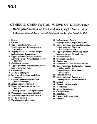
General orientation views of dissection
Midsagittal section of head and neck, right lateral view
A close-up view of the larynx of this specimen is to be found in 83-6.
- Scalp
- Calvaria
- Upper pointer: Falx cerebri Lower pointer: Superior sagittal sinus
- Right pointer: Great cerebal vein Left pointer: Straight sinus
- Upper pointer: Mesencephalon Lower pointer: Cerebral aqueduct
- Confluence of the sinuses
- Upper pointer: Fourth ventricle Lower pointer: Pons
- Cerebellum
- Medulla oblongata
- Margins of foramen magnum
- Cerebellomedullary cistern (cisterna magna)
- Posterior arch atlas
- Upper pointer: Anterior arch atlas Lower pointer: Dens axis
- Spinous process axis
- Intervertebral disc
- Spinal cord
- Laryngeal part of pharynx
- Left pointer: Fornix Right pointer: Corpus callosum
- Upper pointer: Third ventricle Lower pointer: Interpeduncular cistern
- Upper pointer: Optic chiasm Lower pointer: Hypophysis
- Frontal sinus
- Sphenoid sinus
- Nasal cavity
- Nasal septum (partially cut away)
- Upper pointer: Bony auditory tube Lower pointer: Nasal part pharynx
- Hard palate
- Upper pointer: Oral cavity Lower pointer: Soft palate
- Tongue
- Oral part pharynx
- Mandible
- Geniohyoid muscle
- Hyoid bone
- Epiglottis
- Larynx (plica vocalis)
- Trachea